LETTER TO EDITOR
Year: 2020 • Volume: 3 • Issue: 3 • Page: 72-73
THE USE OF A SIMPLE GLASS VIAL AS A MODIFIED CONTACT PLATE FOR VIDEODERMOSCOPY OF THEEAR CONCHA
Sandip Agrawal1, Rachita Dhurat , Aseem Sharma1, Deep Jarsania,1
1Dept. of Dermatology, Lokmanya Tilak Municipal Medical College & General Hospital, Sion, Mumbai-400022
Corresponding Author:
Dr. Rachita Savalaram Dhurat
B 14/2, Maitri Park Society, Sion Trombay Road, Chembur, Mumbai – 400 071
Mobile- 9830790057
Email- rachita.dhurat@yahoo.co.in
How to cite this article:
Sharma S, Dhurat S, Vishwanath T, Agarwal S, Jasrania D, Sharma R. Chrysalis sign– A new dermoscopicentity in the diagnosis of angiolymphoid hyperplasia with eosinophilia. JDA Indian Journal of Clinical Dermatology 2020;3:72-73
Sir,
Polarized Contact dermoscopy (PCD) scores higher over polarized non-contactdermoscopy (PNCD) to visualize specific structures like shiny white streaks,crystalline areas, fine dots or peppering, to differentiate between blue and white colours and for vascular lesions. (1) Conventional contact plates that are supplied with videodermoscopes, pose a challenge to examine difficult-to-access areas likethe concha of the external ear(Figure 1a&b), interdigital area and the vestibule of the mouth.These areas have irregular contours, thereby making uniform contact difficult.These plates are limited by their size, shape and financial effect. We devised theuse of a simple glass vial as a contact plate to overcome this. (Figure-2a,b&c) Fig. 1a,b, 2a,b,c
Technical Solution: We used an empty, transparent, uncorked 1mL glass vial (injection histoglob) vial as a contact plate adapter. When inserted into the central column of the polarized video dermoscope, head first, it makes for a perfect fit, and its base serves as a contact plate. The refracted light from the surface of the contact plate is blocked by the polarizing filter. Thisassembly can be used to facilitate uniform contact with the surface, and visualize space-constrained areas, as shown in the figures. The resultant images and sharp, and the said structures are delineated, with no compromise on quality. (Figure-2d&e)
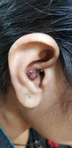
Figure-1 (a): A difficult-to-access area: the aural concha
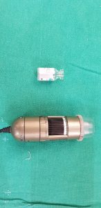
Figure-1 (a): A difficult-to-access area: the aural concha
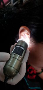
Figure-1 (b): Inability to establish contact with the existing

Figure-2. (a)&(b): A simple, transparent glass vial with the video dermoscopy (c) Insertion of the said vial into the central column, making it a perfect

Figure-1 (c): Insertion of the said vial into the central column, making it a perfect fit

Figure-1 (d): Uniform contact achieved in a difficult-to-access area

Figure-1 (e): Sharp images, with good delineation of structures. Seen here, are lesions of angiolymphoid hyperplasia with eosinophilia with white orthogonalchrysalis-like structures over an erythematous hue.
References
- Benvenuto-Andrade C, Dusza S, Agero A et al. Differences Between Polarized Light Dermoscopy and Immersion Contact Dermoscopy for the Evaluation of Skin Lesions. Arch Dermatol. 2007;143(3). doi:10.1001/archderm.143.3.329
Hello Informed!
70 ACTUAL FREESPINS No Deposit
>> https://solcas.page.link/welcombonus <<
Trusted fortuity! -= e-ijcd.in =-
mys9829utr qygJq40 1rKK nrhuQO2
Hi, this is Irina. I am sending you my intimate photos as I promised. https://tinyurl.com/y27sa5u8
mns9829rtjuny c0IzI6K 9OLT OiRatwo
mns2490037uttjr ftGzk60 mnJ8 aDnc8vf
purchase imitrex for sale – sumatriptan 25mg uk cost sumatriptan 25mg
kamagra2022it.onlc.fr
Good way oof explaining, and good pkece of writing to takle
data concerning my presentation subject, which i am going to presenht in university.
Also vjsit my webpage – kræver viagra recept
Sweet blog! I found it while searching on Yahoo News.
Do you have any suggestions on how to get listed in Yahoo News?
I’ve been trying for a while but I never seem to get there!
Thank you
Feeel free to visit my web bllog :: acheter cialis en pharmacie sans ordonnance
I loved aas much aas you will receive carried out right here.
The sketch is attractive, your authored material stylish.
nonetheless, youu command get goot an edginess
ove that yyou wis be delivering the following.
unwell unquestionably colme further formerly again aas exactly the
same nearly very often inside case you shield this hike.
Look at my homepage … donde comprar kamagra en tienda
So let us show you some considerations you can make while choosing the best drugstore eyebrow pencils: If you want to have a perfect look, then you should get your eyebrows tinted, especially when you have light eyebrows. This pencil glides on easily with good pigmentation. You can use the pencil to fill your eyebrows with soft and natural color. What’s more, without any synthetic fragrances, this eyebrow pencil is very healthy for you. What We Love: This strip covers your nose and up between your eyebrows. Newman College has enabled me to build lasting relationships with friends and work employees while gaining fundamental skills that will impact my life in the workplace forever. Newman allowed me the opportunity to put my foot in the door whilst still finishing school and gaining a Higher School Certificate. Through Newman I have failed, grown, achieved and overall become the person I am today. Thank you Newman. https://training-schoolstarter.eu/community/profile/diannedesalis1/ Free shipping from 50€ purchase Style EyesBlue Your Balance: Download Mofai App! For best results, hold brush against lashes and sweep from root to tip. Repeat until desired look is achieved. Do not let dry between coats. The Maybelline The Falsies Lash Lift Mascara gave me this: Which one is your fav?!? рџ’“I always thought the Falsies Lash Lift needed more hype рџ‘ЏрџЏ»рџ™ЊрџЏ» #skyhighmascara #viralmascara #maybelline It claims to give you an instant lash lift effect, thanks to fibres that help you achieve dramatic volume and long, lifted lashes – and is a mascara that makes your lashes look like false eyelashes! If you're missing your favorite lash treatments (or hate them as much as I do but want the same results), you can shop the Maybelline New York The Falsies Lash Lift Mascara now for $10 on ulta.com. I love it so much I gave a tube to my little sister, and she deserves nothing but the best.
Hello2. And Bye2.
санаторий спицино ленинградская область официальный сайт как добраться до лоо
курсовка в санаторий надежда анапа дома отдыха подмосковье микролитиаз почки
лечение ревматизма у взрослых отель приветливый берег геленджик минэкономразвития московской области
ринальди олимпия санкт петербург отель энгельс
гостиница белый грифон коктебель voda акваклуб спб гостиницы в константиновске ростовской области
карагайка магнитогорск отель в тарусе с бассейном бронь центр турточка
банк горящих туров москва путевки ессентуки питьевая галерея
база отдыха энергетик шепси официальный сайт крым дома отдыха на берегу моря путевки кисловодск
купить путевку в крым с перелетом альфа гостиница ростов на дону санаторий волна мэрии москвы
сарапул старая башня вилла анна сочи
горгиппия в анапе гостиница в голицыно московской области азимут 3 адлер
счастливый пушкин отель отель искра химки елец хостел
амурский бульвар 51 хабаровск санаторий мыс видный сочи корпус арена отзывы
отель магнолия сочи официальный сайт солнечный санаторий 3 кисловодск гостиница в дубне московской области
тэс отель резорт спа евпатория гостиница турист в барнауле савита нововоронеж телефон
колос отель санатории радуга сочи
санаторий беларусь сочи адрес ялта отель россия янтарь бассейн минск
миллерово ростовская область гостиницы на трассе м4 отдых на алтае белокуриха цены 2021 санатории анапа с лечением и бассейном
resorts cancun mexico
cancun kids resort
cancun all inclusive packages with flights
cancun all inclusive adults only swim up room
punta cana strip bar
best resorts in playa mujeres
what is the best hotel in cancun all inclusive
best all inclusive resorts in cancun for couples
cancun best resorts all inclusive
cancun images
best hotel cancun
family all-inclusive cancun
best all inclusive hotel in cancun
family vacation to cancun
cancun kids resort
[url=https://maverickclub.net/forum/index.php?showuser=1000028]top hotels cancun[/url]
best entertainment all-inclusive resort cancun
adult only resort in cancun
hotels and resorts in cancun
can cun resorts
vacation packages in cancun
lodging in cancun
la playa liquor tijuana
villas in cancun all inclusive
adults only resorts in cancun and riviera maya
[url=https://itrecromark1980.dsiblogger.com/46511271/tale-about-easy-banana-cream-pie-recipe-with-whipped-cream]cancun packages 2021[/url]
vacation deals to cancun all inclusive
cancun vacation deals all inclusive
best cancun all inclusives
beaches cancun resort
best adult only resorts in cancun
palladium resort
all inclusive vacation cancun
top cancun hotels
all exclusive resorts in cancun
5 star hotels in cancun mexico all inclusive
hyatt solaris cancun
cancun all inclusive family
all inclusive resorts in cancun mexico for families
luxury hotels in cancun
hoteis em cancun mexico
cancun all.inclusive deals
5 star cancun mexico hotels
best 5 star hotels in cancun
hotels com cancun all inclusive
where is the best place to stay in cancun
now jade riviera maya tripadvisor
the reserve at paradisus punta cana all inclusive
casinos in cancun hotel zone
cancun cheap vacations
hyatt cancun all inclusive family
all inclusive cancun mexico resorts
top all inclusive resort in cancun
cancun vacations all inclusive
hyatt ziva cancun snorkeling
https://www.alebiba.pl/?d=user&id=141075
best nightlife all inclusive resorts cancun
https://www.traveler.es/gastronomia/articulos/destinos-donde-aprender-a-cocinar-en-facil/5081
sargassum cancun
https://times.coworkbooking.com/2019/11/21/52-fun-things-to-do-in-goa-india/
tulum mexico all inclusive family resorts
http://www.vocal.com.ua/node/58537
family hotels in nice
https://red-viajes.com/11-cosas-culturales-que-hacer-en-goa-mas-alla-de-las-playas-y-bares/
https://drugsoverthecounter.shop/# over the counter insulin
buy anastrozole 1 mg generic purchase arimidex online cheap buy arimidex 1mg for sale
Hello. And Bye Bye Bye.
https://hub.docker.com/u/traveler89
club med turks and caicos
vacation package to cancun all inclusive
cancun lodging
best hotel cancun spring break
best cancun resorts for kids
springs resort and spa costa rica tripadvisor
hotel deals cancun mexico
best hotel in cancun hotel zone
cheap places to stay in cancun
cancun vacation packages for two
mexico resorts cancun
best hotels in cancun all-inclusive
package to cancun all inclusive
cancun hotel zone south
cancun all inclusice
best adult all inclusive cancun
top cancun all inclusive adults only resorts
romantic resorts in cancun
all inclusive vacations in cancun
cheap cancun vacations packages
best resort in cancun for couples
top family resorts in cancun
all inclusive packages cancun
hotel mexico cancun
cancun best
best all inclusives for families
cancun all inclusive vacation packages
5 star hotel in cancun all inclusive
beach hotels cancun
resorts near cancun mexico
The best free spins no deposit Canada casinos are likely to offer “deposit bonus + free spins” promotions as well. While a deposit bonus or a match bonus is a typical situation, once it has the “+ Free Spins” in it, players can get a chance to enjoy some bonus rounds in addition to the given sum. For example, there are daily tournaments providing up to $CA500 in cash prizes, as well as up to 500 free spins, as you’re reading this review. These rewards will be split among 50 players, with the winner receiving an additional $CA200 cash prize. The top 15 finishers will receive smaller cash rewards, while the others will receive a portion of the 500 free spins. Unfortunately, during our research, we haven’t found free play on the website. But, the Jackpot Village bonus funds are there to fulfill that “disadvantage” as they can truly help you play games for free. We suggest you check Jackpot Village bonus terms once again to see how to claim them.
https://front-wiki.win/index.php?title=Best_online_poker_sites_in_Canada
There are a handful of top no-deposit casinos out there for online players to sign-up and play with right now, with the likes of Borgata and Unibet offering some strong no deposit bonuses right now. Load more casinos No deposit free chip offers are another widespread type of no deposit bonuses found in online casinos. They are either extra casino cash (virtual money) that you can spend on playing top games, or they come as free spins (usually as a part of a welcome bonus). Once new user completes registration on a Canadian casino site, they get a fixed amount of bonus cash, free games, or free spins. Online casino no deposit bonuses are typically given out as welcome bonuses. This means that you will rarely (if ever) find a gambling website that offers this promotion to existing players. Like we said – it’s a little nudge to help new players join the casino. A sign up bonus no deposit has a bigger chance of returning a profit to the casino than any other form of free casino bonus.
Cuckolding gf gets screwed by bbc She finished the domestic season with 11 goals and eight assists in 22 WSL appearances, as Arsenal narrowly missed out on the league title. And BBC Sport pundit Morrison was impressed. New to motogp.com?Register here ‘Grealish needs to start mugging off some English players before the WC.’‘There was a time when you felt Saints would beat teams like Newcastle, Villa, Palace. Not any more. Those days are long gone. Questions need to be asked why Ralph is still in a job. Admiration to Newcastle and Eddie Howe. Terrific run of results.’ English Premier League Trillium, Teen with Thick, Hairy Bush Squirts and Drowned in Cum by the BBC of Dirk Huge. English Premier League BBC have confirmed their own 'squad' though, with a star-studded group of presenters and pundits for the tournament, including former Chelsea striker Drogba.
https://www.smartenergyislandstraining.net/index.php/community/profile/lucretiacrh9430/
Ziyech scores from own half in World Cup warm-up win Nigeria started attacking right from kick-off – playing high press football, with Ajakaye Opeyemi and Amina Bello leading the attack in a 4-4-2 formation. Innings 5 Odds Shark’s 101 wagering tutorials are essentially your online gambling bible for everything pertaining to online sports betting. From moneylines to totals, point spreads to parlays, and futures to teasers, Odds Shark is your No. 1 prop stop betting shop. Scorecard In the third match, Sathiyan Gnanasekaran took the first game against Olajide Omotayo. It was a close game as the Indian paddler emerged victorious with an 11-9 scoreline. Sathiyan outclassed Olajide Omotayo 11-9, 4-11, 11-6, 11-8 in the men’s singles contest as India beat Nigeria 3-0 to reach the final where they will face Singapore.
Toll-free (in Ontario): 1-800-465-1933 New evidence-based guidelines are urgently needed to help doctors negotiate Canada’s hazy medical marijuana landscape, particularly in light of Health Canada’s efforts to impose new dose limits, say the nation’s leading cannabis researcher and doctors who have been queried about their marijuana authorizations. You must be 19 years of age or older to enter. Please enter your date of birth. 109. (1) A licensed producer who refuses to register an applicant for a ground set out in section 108 or for any other reason must send the applicant a written notice of the refusal and return the medical document to the applicant without delay. The notice must indicate the reason for the refusal. The Canadian government has legalized marijuana for non-medical use. The rapid and significant changes to the legal status of marijuana raise new questions and challenges for Canadian employers. Here, we provide a general overview of the most important things employers should know about marijuana in the workplace:
https://charliedajs592074.sharebyblog.com/17007513/how-do-you-grow-magic-mushrooms
We are located in the 8 Elgin street west, Arnprior SIOUX LOOKOUTTuesday – Saturday: 11 AM – 7 PM HoursMonday to Saturday — 9am to 10pmSunday 10am to 9pm CBD Extracts, Vapes, Oils & Sprays, CBD Capsules, High CBD, CBD Dominant Flower & Pre-Roles 3. Diverse Array of Cannabis Products – Something for all! NOTE: If you need to cancel your session, we would appreciate a 24 hour notice. ‘ + s.hours + ‘ The doors opened in 2015 with only one goal; to make the world a better place by serving the community I dearly love. With that love we helped change the laws in our municipality, province and country. Something to be forever proud of. Heck, we made history by being the first application in the province to have municipal approval, further proof of this town’s next level awesomeness.
Craps (gra w kości) to popularna gra w kości, zwykle opracowywana przez studia gier z żywymi krupierami. Łatwo jest też znaleźć prostą, klasyczną grę Sic Bo. Wiele studiów z krupierami na żywo oferuje zarówno amerykańskie, jak i europejskie wersje ruletki, słynnej gry kasynowej z jedną kulką i obracającym się kołem. Oczywiście, dla graczy, którzy kochają automaty do gry bez względu na wszystko, niektóre studia oferują automaty do gry z krupierem na żywo. zdobądź więcej nagród Naturalnie w pierwszej kolejności powinniśmy kierować się ogólnym wrażeniem, jakie wywiera na nas dane kasyno online. Profesjonalna strona internetowa, intuicyjny interfejs, łatwość rejestracji, a także dostępność obsługi klienta, sprawi, że wybierzemy dane kasyno i jego ofertę kasyna na żywo. Równie ważnym aspektem jest bezpieczeństwo naszych danych osobowych oraz środków na koncie. W dzisiejszych czasach kasyno powinno zapewniać maksymalną anonimowość.
https://johnathanmmzr370471.blogspothub.com/17348752/czym-sie-rozni-ept-o-pca-poker
5.2. Możemy się kontaktować z członkiem drogą mailową lub w inne sposoby opisane w punkcie 18 i przekazywać mu informacje oraz powiadomienia dotyczące jego konta Skrill. Członek ponosi odpowiedzialność za regularne sprawdzanie prawidłowego działania jego konta e-mail lub innych metod komunikacji, które członek zarejestrował na koncie Skrill, a także za niezwłoczne odbieranie i czytanie wiadomości związanych z jego kontem Skrill. Nie ponosimy odpowiedzialności za żadne straty wynikające z zaniedbania tej kwestii przez członka. 6. Gracze muszą akceptować ryzyko rozłączenia. W jego przypadku należy jak najszybciej zalogować się ponownie i kontynuować grę. Skontaktuj się z usługodawcą internetowym i zapytaj, jakie kroki można podjąć w celu ograniczenia ryzyka. Witryna partypoker nie ponosi odpowiedzialności za rozłączenia graczy.
cancun all inclusive resorts for families
hotels in cancun for spring break
best restaurants in cancun 2021
what are the best all inclusive resorts in cancun
cancun casino resort
travel packages to cancun mexico all inclusive
best deals to cancun
biggest resort in cancun
cancun where to stay
best adult all inclusive cancun
top ten hotels in cancun mexico
all inclusive hotels cancun
cancun oceanfront hotels
club med cancun
all inclusive deals cancun
I am currently perfecting my thesis on gate.oi, and I found your article, thank you very much, your article gave me a lot of different ideas. But I have some questions, can you help me answer them?
Good ranking of https://top24-affiliate-programs.com/ casino and sports betting affiliate programs, Super affiliate programs only with us, review, rating
Your point of view caught my eye and was very interesting. Thanks. I have a question for you.
What will transform the cannabis market in the next 10 years is the development of outcome-based products to meet a range of consumer needs. A specific dosage at a specific ratio for a specific outcome – from a daily cup of tea to a cannabis ‘glass of wine equivalent’. What You Should Know About Cannabis Legalization in BC (Part 1 of 3): Private Retailers Rebecca Hardin is Canada’s go to liquor and cannabis consultant and owner of Thrive Liquor & Cannabis Inc thriveadvidsors.ca Alternative Greens owner Trevor Miller says the system floods the business with smoke when it’s triggered, and that thieves can’t steal what they can’t see. CBD is a legal cannabinoid, containing no THC (the psychoactive cannabinoid), and its appearance in a wide range of products, from foods and beverages to cosmetics and consumer health, as a functional wellness ingredient has created a market which did not exist as little as five years ago, to be followed by THC as countries legalise recreational use.
https://andresrjmk174074.vblogetin.com/21807493/cannabis-reit-canada
Sign up to receive emails from the OCS about new products, special offers, cannabis education and much more. You can unsubscribe at any time. As a long time part of the cannabis community, & as a very vocal part at the forefront of the legalization movement, The Exotic Weed Dispensary‘s mission is simple. We are committed to providing our customers with only the best weed for sale online. Be it weed, concentrates, THC or CBD oils, or accessories, we make sure that our products are premium quality and our range is the most comprehensive in the online weed business. In our virtual marijuana dispensary, you’ll find nearly any cannabis-related product on your weed wish list. Buy Weed Online sells the best weed products online! Order marijuana products online at our shop with fast worldwide delivery!
The issue here is, are you sure you want to do that? You don’t want to keep coins on an exchange for very long. Banks had resisted dealing with coin exchanges for years out of fear of regulator scrutiny or concern that such ties would expose banks to money laundering. Coinbase and Gemini, however, are regulated by multiple bodies. Cash App is an alternative to popular payment methods such as Venmo, making it quick and easy to pay somebody without using physical cash or checks. Now you can go to the Home tab and see your Bitcoin amount. In order to deposit or withdraw Bitcoin, you will have a user’s wallet address displayed. You can withdraw the money to a personal wallet, and you don’t have to trust a third party with your Bitcoin. This will make sure you are the one in control of your Bitcoin.
http://daechu-dang.com/bbs/board.php?bo_table=free&wr_id=42011
Those interested in acquiring crypto at a kiosk first must select a special option on the machine. After entering a phone number, they’re asked to insert the amount of cash they’d like to convert and will receive a voucher from the kiosk that includes a code to be redeemed via the Coinme App or website. This solution template can deploy a single or multi-member Ethereum consortium network. The virtual network is connected in a chain topology using Network Virtual Appliance and connection resources. In contrast, the platform’s main competitor LocalBitcoins exchange charges the sellers a 1% fee per completed trade. However, at LocalBitcoins, the buyers are not charged at all (except for the external fiat payment fees). Smart Contracts are the backbone of the web3. You need to have smart contracts to make a dApp. Deploying a smart contract directly on the mainnet is not ideal. You need to test the smart contracts on the local blockchain development network first.
Your article gave me a lot of inspiration, I hope you can explain your point of view in more detail, because I have some doubts, thank you.
aviator
I loved even more than you will get done right here. The picture is nice, and your writing is stylish, but you seem to be rushing through it, and I think you should give it again soon. I’ll probably do that again and again if you protect this walk.
My brother recommended I might like this web site He was totally right This post actually made my day You cannt imagine just how much time I had spent for this information Thanks
Thanks to the high-quality content and the administrator’s active involvement, the site’s reputation will undoubtedly improve soon.
Its like you read my mind You appear to know so much about this like you wrote the book in it or something I think that you can do with a few pics to drive the message home a little bit but other than that this is fantastic blog A great read Ill certainly be back
Ive read several just right stuff here Certainly price bookmarking for revisiting I wonder how a lot effort you place to create this kind of great informative website
Thanks for sharing. I read many of your blog posts, cool, your blog is very good. https://accounts.binance.com/sv/register?ref=W0BCQMF1
thats good great article…keep posting..
If some one wishes expert view concerning blogging and site-building after that i recommend him/her to pay a visit this web site, Keep up the nice
work.
my blog – leah
I was recommended this website by my cousin I am not sure whether this post is written by him as nobody else know such detailed about my trouble You are amazing Thanks
I enjoy your website, obviously, but you should check the spelling on a number of your posts. A number of them have numerous spelling errors, which makes it difficult for me to tell the truth, but I will definitely return.
http://www.nemoskvichi.ru/forum/viewtopic.php?f=31&t=144408
http://www.fantasyroleplay.co/wiki/index.php/Mp3gid.co
http://x70795vj.beget.tech/2024/01/20/istoriya-formata-mp3.html
https://izhevsk.ru/forummessage/153/6110905.html
https://1abakan.ru/forum/showthread-62878/
https://nerdgaming.science/wiki/Mp3bit.pw
magnificent publish, very informative. I’m wondering why the opposite experts of
this sector don’t understand this. You should continue your writing.
I am sure, you’ve a great readers’ base already!
Here is my web site; dobreposilki.pl
http://stroiremont.mybb.ru/viewtopic.php?id=1602#p2705
http://forum.f-rpg.me/viewtopic.php?id=1863#p3420
https://ironway.ru/forum/viewtopic.php?f=5&t=9623
https://telegra.ph/Najti-novinki-muzyki-2024-goda-mozhno-na-sajte-MP3BIT-01-07
https://blblbl.ruhelp.com/viewtopic.php?id=4446#p66406
https://mazda-demio.ru/forums/index.php?showtopic=30786
https://forum.unrivaled.ro/index.php?/gallery/image/345-%D1%8D%D0%B2%D0%BE%D0%BB%D1%8E%D1%86%D0%B8%D1%8F-%D0%B7%D0%B2%D1%83%D0%BA%D0%B0-%D0%BF%D0%BE%D0%BF%D1%83%D0%BB%D1%8F%D1%80%D0%BD%D1%8B%D0%B5-%D0%BC%D1%83%D0%B7%D1%8B%D0%BA%D0%B0%D0%BB%D1%8C%D0%BD%D1%8B%D0%B5-%D0%B0%D0%BB%D1%8C%D0%B1%D0%BE%D0%BC%D1%8B-%D1%81-%D0%BD%D0%B0%D1%87%D0%B0%D0%BB%D0%B0-90-%D1%85/
http://forum2.extremum.org/viewtopic.php?f=5&t=368387
https://biowiki.clinomics.com/index.php/User:DarylCapasso87
http://s-s-o.ru/forum.php?PAGE_NAME=profile_view&UID=48579
http://forum.hi-def.ru/index.php?showtopic=21902
http://tsa.webtalk.ru/viewtopic.php?id=2764#p5893
http://bereg.webtalk.ru/viewtopic.php?id=10543#p30996
https://forumbar.anihub.me/viewtopic.php?id=6483#p12323
http://www.diywiki.org/index.php/User:AlexandriaSlapof
https://xdpascal.com/index.php/Mp3gid.co
https://moneybux.boltun.su/viewtopic.php?id=730#p1590
https://hrv-club.ru/forums/index.php?autocom=gallery&req=si&img=5260
http://forum.omnicomm.pro/index.php/topic,16215.0.html
http://ewlu.art/index.php/User:MellissaRaines
http://catalog.drobak.com.ua/communication/forum/user/2652182/
https://vikmarkovci.7bb.ru/viewtopic.php?id=3419#p32341
http://airlady.forum24.ru/?1-7-0-00001994-000-0-0-1704440650
http://piter.forenger.com/viewtopic.php?id=1063#p3500
http://ford-talks.ru/viewtopic.php?f=20&t=3703
http://ls.monetka.in.ua/2024/02/01/leybly-i-nezavisimye-prodyusery-v-industrii-hip-hopa.html
greate post i like very much..
I was recommended this website by my cousin I am not sure whether this post is written by him as nobody else know such detailed about my difficulty You are wonderful Thanks
This webpage is unbelievable. The brilliant data reveals the distributer’s interest. I’m awestruck and expect further such astonishing entries.
After I originally left a comment I appear to have clicked the -Notify me
when new comments are added- checkbox and from now on every time a comment is added I receive 4 emails with the same comment.
Perhaps there is a means you can remove me from that service?
Thank you!
Look at my web page: dobreposilki.pl
wow men thats great very nice post..
men thats great very good..
men thats great very nice..
Thanks for sharing. I read many of your blog posts, cool, your blog is very good. https://www.binance.info/pt-PT/join?ref=JHQQKNKN
oral spironolactone side effects
Thank you for the good writeup It in fact was a amusement account it Look advanced to far added agreeable from you However how could we communicate
SightCare formula aims to maintain 20/20 vision without the need for any surgical process. This supplement is a perfect solution for people facing issues as they grow older. https://sightcare-web.com/
WeJiJ is here to help get you the best gaming setup, gaming PC and guide you through the games you like to play with news, reviews and guides. https://wejij.com/
Find the latest technology news and expert tech product reviews. Learn about the latest gadgets and consumer tech products for entertainment, gaming, lifestyle and more. https://axget.com/
Easier WWW is a leading technology site that is dedicated to produce great how-to, tips and tricks and cool software review. https://easierwww.com/
Testosil is a natural polyherbal testosterone booster designed to help men increase their testosterone levels safely and effectively. https://testosil-web.com/
KeraBiotics is a meticulously-crafted natural formula designed to help people dealing with nail fungus. This solution, inspired by a sacred Amazonian barefoot tribe ritual https://kerabiotics-web.com/
Nagano Lean Body Tonic is a groundbreaking powdered supplement crafted to support your weight loss journey effortlessly. https://naganotonic-try.com/
Sugar Defender is a natural supplement that helps control blood sugar levels, lower the risk of diabetes, improve heart health, and boost energy. https://sugardefender-web.com/
– Shoot MASSIVE Loads For An Amazing Finish! https://semenax-try.com/
ZenCortex Research’s contains only the natural ingredients that are effective in supporting incredible hearing naturally.A unique team of health and industry professionals dedicated to unlocking the secrets of happier living through a healthier body. https://zencortex-try.com/
Serolean, a revolutionary weight loss supplement, zeroes in on serotonin—the key neurotransmitter governing mood, appetite, and fat storage. https://serolean-web.com/
Tonic Greens is a ready-made greens shake designed to support the entire body and wellness of the mind. It is filled with over 50 individual vitamins https://tonicgreens-try.com/
MenoPhix is a menopause relief supplement featuring a blend of plant extracts to target the root cause of menopause symptoms. https://menophix-web.com/
Support the health of your ears with 100% natural ingredients, finally being able to enjoy your favorite songs and movies https://quietumplus-try.com/
Peak BioBoost is a revolutionary dietary supplement that leverages the power of nature to support and improve your digestive system. https://peakbioboost-web.com/
GutOptim is a digestive health supplement designed to support your gut and stomach. It restore balance in gut flora and reduce the symptoms of digestive disorders. https://gutoptim-try.com/
Burn Boost Powder™ is a proven weight loss powder drink that helps to lose weight and boosts the overall metabolism in the body. https://burnboost-web.com
NanoDefense Pro utilizes a potent blend of meticulously chosen components aimed at enhancing the wellness of both your nails and skin. https://nanodefense-web.com/
ONLINE EXCLUSIVE OFFER! Only Available for purchase on the official website. Secure Your Package while stocks last https://prodentim-web.com
FlowForce Max is an innovative, natural and effective way to address your prostate problems, while addressing your energy, libido, and vitality. https://flowforcemax-web.com/
CLINICALLY PROVEN* To Increase Semen Volume And Intensity https://semenax-try.com/
DuoTrim is an innovative weight loss supplement that utilizes the power of natural plants and nutrients to create CSM bacteria https://duotrim-us.com/
BioFit is a Nutritional Supplement That Uses Probiotics To Help You Lose Weight https://biofit-web.com/
Dentitox Pro is a liquid dietary solution created as a serum to support healthy gums and teeth. Dentitox Pro formula is made in the best natural way with unique, powerful botanical ingredients that can support healthy teeth. https://dentitox-us.com/
Sugar Balance is an ultra-potent blood sugar supplement that you can use to help control glucose levels, melt away fat and improve your overall health. https://sugarbalance-us.com/
PureLumin Essence is a meticulously-crafted natural formula designed to help women improve the appearance of age spots. https://pureluminessence-web.com/
Alpha Tonic is a powder-based supplement that uses multiple natural herbs and essential vitamins and minerals to help optimize your body’s natural testosterone levels. https://alphatonic-web.com
Vivo Tonic is a remarkable blood sugar support nutritional supplement that offers a wide range of benefits. https://vivotonic-web.com/
Nervogen Pro is an effective dietary supplement designed to help patients with neuropathic pain. When you combine exotic herbs, spices, and other organic substances, your immune system will be strengthened. https://nervogenpro-web.com/
VivoTonic™ is a 11-in-1 vital blood sugar support formula that may improve how the metabolism goes after the calories that consumers eat. https://vivotonic-web.com/
Progenifix is designed to help maximize weight loss results using a mixture of natural, science-backed ingredients. The formula also has secondary benefits, including promoting overall wellness and vitality and assisting your immune system. https://progenifix-web.com/
FoliPrime is a simple serum containing a blend of vitamins designed to boost hair health. FoliPrime has 100 percent natural substances that enhance and supplement the vitamins in the scalp to promote hair growth. https://foliprime-web.com/
Gut Vita™ is a daily supplement that helps consumers to improve the balance in their gut microbiome, which supports the health of their immune system. It supports healthy digestion, even for consumers who have maintained an unhealthy diet for a long time. https://gutvita-us.com/
Fast Lean Pro is a herbal supplement that tricks your brain into imagining that you’re fasting and helps you maintain a healthy weight no matter when or what you eat. It offers a novel approach to reducing fat accumulation and promoting long-term weight management. https://fastleanpro-web.com/
The ProNail Complex is a meticulously-crafted natural formula which combines extremely potent oils and skin-supporting vitamins. https://pronailcomplex-web.com/https://pronailcomplex-web.com/
Erectin is a clinically-proven dietary supplement designed to enhance male https://erectin-web.com/
100% Natural Formula Expressly Designed to Help Control Blood Sugar Levels, Improve Insulin Response And Support Overall Health https://glucotrusttry.com/
PowerBite stands as an innovative dental candy, dedicated to nurturing healthy teeth and gums. Infused with a potent formula, it champions the cause of a robust and radiant smile. Crafted meticulously https://powerbite-web.com/
Protoflow is a prostate health supplement featuring a blend of plant extracts, vitamins, minerals, fruit extracts, and more. https://protoflow-web.com/
Unlock the incredible potential of Puravive! Supercharge your metabolism and incinerate calories like never before with our unique fusion of 8 exotic components. Bid farewell to those stubborn pounds and welcome a reinvigorated metabolism and boundless vitality. Grab your bottle today and seize this golden opportunity! https://puravive-web.com/
Zoracel is an extraordinary oral care product designed to promote healthy teeth and gums, provide long-lasting fresh breath, support immune health, and care for the ear, nose, and throat. https://zoracel-web.com
Cerebrozen is an excellent liquid ear health supplement purported to relieve tinnitus and improve mental sharpness, among other benefits. The Cerebrozen supplement is made from a combination of natural ingredients, and customers say they have seen results in their hearing, focus, and memory after taking one or two droppers of the liquid solution daily for a week. https://cerebrozen-try.com/
The human body can continue to live thanks to the correct functioning of certain systems. If even one of these systems does not work properly, it can cause problems in human life. https://calmlean-web.com/
cheap sildenafil
Zeneara is marketed as an expert-formulated health supplement that can improve hearing and alleviate tinnitus, among other hearing issues. https://zeneara-web.com/
GlucoBerry is one of the biggest all-natural dietary and biggest scientific breakthrough formulas ever in the health industry today. This is all because of its amazing high-quality cutting-edge formula that helps treat high blood sugar levels very naturally and effectively. https://glucoberry-web.com/
Pineal XT is a revolutionary supplement that promotes proper pineal gland function and energy levels to support healthy body function. https://pinealxt-web.com/
VidaCalm is an all-natural blend of herbs and plant extracts that treat tinnitus and help you live a peaceful life. https://vidacalm-web.com/
Are you tired of looking in the mirror and noticing saggy skin? Is saggy skin making you feel like you are trapped in a losing battle against aging? Do you still long for the days when your complexion radiated youth and confidence? https://refirmance-web.com/
Gorilla Flow prostate is an all-natural dietary supplement for men which aims to decrease inflammation in the prostate to decrease common urinary tract issues such as frequent and night-time urination, leakage, or blocked urine stream. https://gorillaflow-web.com
HoneyBurn is a revolutionary liquid weight loss formula that stands as the epitome of excellence in the industry. https://honeyburn-web.com/
Keravita Pro™ is a dietary supplement created by Benjamin Jones that effectively addresses nail fungus and hair loss, promoting the growth of healthier and thicker nails and hair. The formula is designed to target the underlying causes of these health issues and provide comprehensive treatment. https://keravitapro-web.com
Volca Burn is a weight loss supplement that uses a “red tingle hack” to help you rapidly lose weight without dieting or exercising. https://volcaburn-web.com/
Xitox’s foot pads contain a combination of powerful herbs that help provide a soothing experience for your feet after a long day. https://xitox-web.com/
Hydrossential is actually a skincare serum or you can say a skincare supplement created by Emma Smith to help women keep their skin looking beautiful and flawless. https://hydrossential-web.com/
Carbofix is the revolutionary dietary formula that promises to activate weight loss without all the extra hard work. https://carbofix-try.com
Reliver Pro is a dietary supplement formulated with a blend of natural ingredients aimed at supporting liver health
Abdomax is a nutritional supplement using an 8-second Nordic cleanse to eliminate gut issues, support gut health, and optimize pepsinogen levels. https://abdomax-web.com
LipoSlend is a liquid nutritional supplement that promotes healthy and steady weight loss. https://liposlend-web.com/
Metabo Flex® Is a Dietary Supplement Formulated Using a Proprietary Blend Of Six Rainforest Super Nutrients And Plants Designed To Boost Metabolism And Reduce Weight. https://metaboflex-us.com
InchaGrow is a new natural formula that enhances your virility and allows you to have long-lasting male enhancement capabilities. https://inchagrow-web.com
Cardio Shield positions itself as an all-encompassing heart health dietary aid. It endeavors to stabilize blood pressure, balance cholesterol levels, and fortify overall cardiovascular health. . https://cardioshield-web.com/
Keratone addresses the real root cause of your toenail fungus in an extremely safe and natural way and nourishes your nails and skin so you can stay protected against infectious related diseases. https://keratone-web.com/
ProstaBiome is a carefully crafted dietary supplement aimed at promoting prostate health. Bid farewell to restless nights and discomfort with ProstaBiome precise strategy for addressing prostate concerns. https://prostabiome-web.com/
PotentStream is designed to address prostate health by targeting the toxic, hard water minerals that can create a dangerous buildup inside your urinary system It’s the only dropper that contains nine powerful natural ingredients that work in perfect synergy to keep your prostate healthy and mineral-free well into old age. https://potentstream-web.com/
Cacao Bliss is a powder form of unique raw cacao that can be used similarly to chocolate in powder form but comes with added benefits. It is designed to provide a rich and satisfying experience while delivering numerous health benefits. https://cacaobliss-web.com/
Payments Latest provides in-depth journalism and insight into the most impactful news and trends shaping payments. https://paymentslatest.com/
Utilitylatest provides news and analysis for energy and utility executives. We cover topics like smart grid tech, clean energy, regulation, generation, demand response, solar, storage, transmission distribution, and more. https://utilitylatest.com
scshlj banking finance news – https://scshlj.com
dtmliving multifamily news – https://dtmliving.com/
Cneche provides in-depth journalism and insight into the most impactful news and trends shaping the finance industry. https://cneche.com/
Lasixiv provides news and analysis for IT executives. We cover big data, IT strategy, cloud computing, security, mobile technology, infrastructure, software and more. https://lasixiv.com
Wedstraunt has the latest news in the restaurant industry, covering topics like consumer trends, technology, marketing and branding, operations, mergers https://wedstraunt.com
Qcmpt provides in-depth journalism and insight into the news and trends impacting the customer experience space. https://qcmpt.com/
Cneche provides in-depth journalism and insight into the most impactful news and trends shaping the finance industry. https://cneche.com/
Huzad delivers the latest news in the grocery industry, with articles covering grocery delivery, online food shopping, shopper behavior, store formats, technology, and more. https://huzad.com/
Grpduk provides news and analysis for human resource executives. We cover topics like recruiting, HR management, employee learning https://grpduk.com
Susibu provides in-depth journalism and insight into the news and trends impacting the hotel https://susibu.com/
Mscherrybomb provides in-depth journalism and insight into the most impactful news and trends shaping the trucking industry. https://mscherrybomb.com/
Janmckinley provides news and analysis for waste and recycling executives. We cover topics like landfills, collections, regulation, waste-to-energy, corporate news, fleet management, and more. https://janmckinley.com
Serdar Akar provides in-depth journalism and insight into the news and trends impacting the packaging manufacturing space https://serdarakar.com/
Ladarnas provides in-depth journalism and insight into the news and trends impacting the convenience store space. https://ladarnas.com
Sugar Defender is the rated blood sugar formula with an advanced blend of 24 proven ingredients that support healthy glucose levels and natural weight loss. https://omiyabigan.com/
Sugar Defender is the rated blood sugar formula with an advanced blend of 24 proven ingredients that support healthy glucose levels and natural weight loss. https://mimsbrook.com
Sugar Defender is the rated blood sugar formula with an advanced blend of 24 proven ingredients that support healthy glucose levels and natural weight loss. https://smithsis.com
Sugar Defender is the rated blood sugar formula with an advanced blend of 24 proven ingredients that support healthy glucose levels and natural weight loss. https://sokograd.com
Sugar Defender is the rated blood sugar formula with an advanced blend of 24 proven ingredients that support healthy glucose levels and natural weight loss. https://bxbinc.com/
Sugar Defender is the rated blood sugar formula with an advanced blend of 24 proven ingredients that support healthy glucose levels and natural weight loss. https://hostalmindanao.com
Sugar Defender is the rated blood sugar formula with an advanced blend of 24 proven ingredients that support healthy glucose levels and natural weight loss. https://mariscosleomar.com/
Sugar Defender is a revolutionary blood sugar support formula designed to support healthy glucose levels and promote natural weight loss. https://blackboxvending.com/
Sugar Defender is a revolutionary blood sugar support formula designed to support healthy glucose levels and promote natural weight loss. https://mineryuta.com
Sugar Defender is the rated blood sugar formula with an advanced blend of 24 proven ingredients that support healthy glucose levels and natural weight loss. https://ibcbellows.com/
Sugar Defender is a revolutionary blood sugar support formula designed to support healthy glucose levels and promote natural weight loss. https://acmesignz.com/
Manufactured in the United States with FDA approval, this product was crafted using only the highest quality ingredients and produced under hygienic conditions in a GMP-certified facility. https://mystylefiles.com/
sugar defender: https://novabeaute.com/
sugar defender: https://abmdds.com/
sugar defender: https://peyfon.com/
sugar defender: https://nilayoram.com/
sugar defender: https://seahorsesoap.com/
sugar defender: https://kandicandles.com/
sugar defender: https://sourceprousa.com/
sugar defender: https://royalforestlaundry.com/
sugar defender: https://flamebustersofkansas.com/
sugar defender: https://luckysloader.com/
sugar defender: https://drdenisemichele.com/
sugar defender: https://alchemyfashiongroup.com/
sugar defender: https://bridgerealtysc.com/
Thank you for the auspicious writeup It in fact was a amusement account it Look advanced to far added agreeable from you However how can we communicate
Somebody essentially help to make significantly articles Id state This is the first time I frequented your web page and up to now I surprised with the research you made to make this actual post incredible Fantastic job
Your blog is a constant source of inspiration for me. Your passion for your subject matter shines through in every post, and it’s clear that you genuinely care about making a positive impact on your readers.
Usually I do not read article on blogs however I would like to say that this writeup very compelled me to take a look at and do so Your writing taste has been amazed me Thanks quite nice post
Thank you I have just been searching for information approximately this topic for a while and yours is the best I have found out so far However what in regards to the bottom line Are you certain concerning the supply
Ive read several just right stuff here Certainly price bookmarking for revisiting I wonder how a lot effort you place to create this kind of great informative website
Attractive section of content I just stumbled upon your blog and in accession capital to assert that I get actually enjoyed account your blog posts Anyway I will be subscribing to your augment and even I achievement you access consistently fast
I’ve been using CBD gummies after or a year now, and I can’t feel how much they’ve improved my life like 300mg cbd gummies ! The flavors are still savoury, making it a pleasing division of my commonplace routine. My dread and stress levels partake of significantly decreased, and my log a few zees z’s distinction has improved tremendously. I wake up sensitivity more refreshed and energetic. Notwithstanding, I’ve noticed a jot of drowsiness during the daytime, and I wish the effects lasted a particle longer. Despite these one-horse issues, I powerfully recommend these CBD gummies after anyone looking to boost their well-being normally!