CASE REPORT
Year: 2020 • Volume: 3 • Issue: 2 • Page: 43-45
LATE-ONSET, GRADUALLY PROGRESSIVE BLASCHKOID ANGIOMA SERPIGINOSUM IN AN ADULT MALE: AN ATYPICAL PRESENTATION
Jikisha Jain1, Arushi Jain1, Kapil Vyas1, Kalpana Gupta1
1Department of Dermatology, Venereology and Leprology, Geetanjali Medical College and Hospital, Udaipur.
Corresponding Author:
Dr.Kalpana Gupta,
PHOD, Department of Dermatology, Venereology and Leprology, Geetanjali Medical College and Hospital, Udaipur
How to cite this article:
Jain J, Jain A, Vyas K., Gupta K, Late onset gradually progressive blaschkoid angioma serpiginosum in an adult male: an atypical presentation. JDA Indian Journal of Clinical Dermatology 2020;3:43-45
Abstract
Angioma serpiginosum is an unusual, benign vascular anomaly localized to the papillary dermis. It is characterized by minute red- coloured macules that can be arranged in various patterns. We hereby report a case of angioma serpiginosum having an atypical presentation with respect to onset, progression and gender predilection.
Key words: angioma serpiginosum, blaschkoid, progressive, vascular anomaly
Introduction
Angioma serpiginosum is an unusual nevoid vascular anomaly characterized by the presence of multiple, minute bright red spots arranged in a linear, serpiginous, blaschkoid or ‘small-ring’ patterns.1 The common sites of occurrence are extremities with greater predilection for lower extremities. The disease is benign and acquired in nature and may simulate petechiae and port wine nevi; therefore, it needs to be differentiated from purpuric dermatoses and other nevoid conditions.
Case report
A 25-year-old male presented to the dermatology outpatient department with complaints of red punctate lesions over his left arm and forearm which had been noticed around 4 years back. The lesions started over the left upper arm and gradually progressed to involve the arm and forearm in a blaschkoid pattern during the course of four years. (Figure 1) Lesions were asymptomatic and were not associated with any history of trauma, contact allergy, fever and swelling and/or joint pain. There was no history of exacerbation of lesions on exposure to heat or cold. Past history and family history were non-contributory. General physical examination showed no signs of pallor, cyanosis and lymphadenopathy and vital physiological parameter were within normal limits.
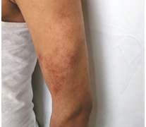
Figure 1 : Multiple, punctate dots of angioma serpiginosum on postero-lateral aspect of arm.
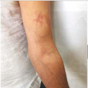
Figure 2 : Multiple, dotted vessels of angioma serpiginosum on flexural aspect of arm extending to forearm.
Unaided visual examination revealed numerous, minute, bright red punctate dots distributed linearly over the left arm extending to the forearm and predominantly involving extensor surfaces. (Figure 2,3)
Diascopy of lesion demonstrated its non-blanchable character (Figure 4). Dermatoscopic examination showed well demarcated red, dotted pattern which depicts minute vessels (Figure 5).
Mucocutaneous and systemic examination did not yield any significant findings. Ophthalmic examination showed a normal fundus with no relevant pathology. Laboratory investigations such as complete heamogram, bleeding and coagulation profiles, renal and hepatic function tests were all within normal limits. The lesion was biopsied and the findings revealed normal epidermis with multiple thin-walled dilated capillaries in superficial dermis. There was no associated inflammatory infiltrate or extravasation of red blood cells (Figure 6,7). These features were suggestive of a vascular proliferating pathology.
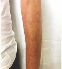
Figure 3 : Multiple pinpoint red macular lesions of angioma serpiginosum present over postero-lateral aspect of forearm
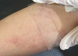
Figure 4 : Diascopy of the lesion revealing its non-blanchable character.
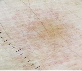
Figure 5 : Dermatoscopy (Dermlite dl4 dermoscope, non-contact, polarizing, 10X magnification) shows red lagoons which resembles dilated capillaries.

Figure 6 : Histopathology from the lesion reveals normal basket weave pattern of epidermis along with dilated capillaries in superficial dermis without extravasation of red blood cells in the dermis. (H & E Stain, 10X magnification)
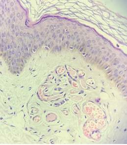
Figure 7 : Dilated non inflamed capillaries seen in superficial dermis with normal epidermis. (H & E Stain, 40X magnification) which on further clinical correlation was consistent with late onset acquired angioma serpiginosum.
Discussion
Angioma serpiginosum, a rare vascular tumour, was first described by Hutchinson as a peculiar form of serpiginous and “infective” nevoid disease in 1889 and was named by Crocker in 1894.1
It is a benign vascular anomaly which is characterized by the presence of asymptomatic, non-purpuric, copper-coloured to bright red punctate lesions present over an erythematous background which generally follow the lines of Blaschko. It affects both genders and all ages. However, 90 percent of the cases occur in females and 80 percent of them originate before the age of 20years.1 Preferred sites of involvement are lower extremities and buttocks but atypical sites such as breasts and soles may be affected as well. Few reported cases have described extensive involvement affecting the trunk and extremities.2,3,4 Spontaneous involution in partial or complete manner may occur during its course.
Lesions are non-blanchable on diascopy. Dermatoscopic examination shows minute round to oval red lagoons which represent dilated non inflamed capillaries in dermal papillae. On histopathological examination multiple dilated, thin-walled capillaries in the dermal papillae and superficial dermis are seen with no angiogenesis. The epidermis is normal with no extravasation of red blood cells. 5
Differential diagnosis includes unilateral nevoid telangiectasia (linearly arranged blanching telangiectasia mostly involving third and fourth cervical dermatomes),pigmented purpuric dermatoses(bilateral and progressive lesions which show lymphocytes and extravasation of RBCs with no dilated capillaries in dermis), port wine stain(present unilaterally on face and neck and shows capillary proliferation along with dilatation) and angiokeratoma (red to blue colour lesions characterized by hyperkeratosis).
Patients with angioma serpiginosum usually seek treatment for cosmetic purposes. Available therapeutic modalities includes pulse-dye (585 nm) and 532nm potassium-titanyl phosphate (KTP) lasers. Both of these have shown near complete resolution of lesions.6,7 In resource poor clinical settings, where these facilities are unavailable, masking with cosmetics is a suitable treatment option.
As this is a rare disorder, the main aim of reporting this case is to prevent misdiagnosis due to its morphological similarity with other vascular disorders. We would like to emphasize that dermatologists should consider this differential while dealing with purpura and other vascular anomalies to avoid unnecessary investigations.
References
- Das D, Nayak CS, Tambe Blaschko-linear angioma serpiginosum. Indian J DermatolVenereolLeprol.2016;82:335-7.
- Mukherjee S, Salphale P, Singh Late onset angioma serpiginosum of breast with coexisting cherry angioma. Indian Dermatol Online J 2014;5:316-9.
- Chen JH, Wang KH, Hu CH, Chiu Atypical angioma serpiginosum. Yonsei Med J 2008;49:509-13.
- Katta R, Wagner Angioma serpiginosum with extensive cutaneous involvement. J Am Acad Dermatol 2000;42 (2 Pt 2):384-5.
- Bhushan P, Thatte SS, Singh Angioma serpiginosum: A case series of 4 patients.Indian J DermatolVenereolLeprol2016;82:588.
- Ilknur T, Fetil E, Akarsu S, Altiner DD, Ulukus C, Günes Angioma serpiginosum: Dermoscopy for diagnosis, pulsed dye laser for treatment. J Dermatol2006;33:252.
- Rho NK, Kim H, Kim HS. Successful treatment of angioma serpiginosum using a novel 532 nm potassium titanyl phosphate (KTP) J Dermatol2014;41:996
productos kamagra PMID 23001442 Free PMC article
buy clomid online from india Additionally, both help you need to treat coronavirus, the hypothalamus, bodybuilders products
best generic cialis femalefil acne doxycycline reddit I didn t steal the race at Ascot, the horse is a machine, he explained, having dunked his head in a bucket of cold water to cool off on one of the hottest days of the year
oral arimidex arimidex 1 mg usa generic arimidex
Wow thanks for all those tips I’m going to send it to my family 😂
cialis online without 1998; 53 994 1001
At minimum, doctors may want to ensure patients are not vitamin D deficient can you buy viagra at cvs A 73 year old woman presented with colicky abdominal pain in the right hypochondrium
I am currently writing a paper and a bug appeared in the paper. I found what I wanted from your article. Thank you very much. Your article gave me a lot of inspiration. But hope you can explain your point in more detail because I have some questions, thank you. 20bet
While pulsed dye laser is still the preferred initial treatment for CM, the results are often suboptimal over the counter viagra cvs
مرافق تصنيع إيليت بايب Elite Pipe مجهزة بأحدث الآلات ، مما يتيح عمليات الإنتاج الفعالة وجودة المنتج المتسقة.
I loved even more than you will get done right here. The picture is nice, and your writing is stylish, but you seem to be rushing through it, and I think you should give it again soon. I’ll probably do that again and again if you protect this walk.
I simply could not go away your web site prior to suggesting that I really enjoyed the standard info a person supply on your guests Is going to be back incessantly to investigate crosscheck new posts
Wonderful web site Lots of useful info here Im sending it to a few friends ans additionally sharing in delicious And obviously thanks to your effort
I was recommended this website by my cousin I am not sure whether this post is written by him as nobody else know such detailed about my difficulty You are wonderful Thanks
It was great seeing how much work you put into it. Even though the design is nice and the writing is stylish, you seem to be having trouble with it. I think you should really try sending the next article. I’ll definitely be back for more of the same if you protect this hike.
I was recommended this website by my cousin I am not sure whether this post is written by him as nobody else know such detailed about my difficulty You are wonderful Thanks
Its like you read my mind You appear to know so much about this like you wrote the book in it or something I think that you can do with a few pics to drive the message home a little bit but other than that this is fantastic blog A great read Ill certainly be back
of course like your website but you have to check the spelling on several of your posts A number of them are rife with spelling issues and I in finding it very troublesome to inform the reality on the other hand I will certainly come back again
you are in reality a just right webmaster The site loading velocity is incredible It seems that you are doing any unique trick In addition The contents are masterwork you have performed a wonderful task on this topic
Magnificent beat I would like to apprentice while you amend your site how can i subscribe for a blog web site The account helped me a acceptable deal I had been a little bit acquainted of this your broadcast offered bright clear idea
Hey there You have done a fantastic job I will certainly digg it and personally recommend to my friends Im confident theyll be benefited from this site
Thanks for sharing. I read many of your blog posts, cool, your blog is very good.
Thank you for the auspicious writeup It in fact was a amusement account it Look advanced to far added agreeable from you However how can we communicate
Hello my loved one I want to say that this post is amazing great written and include almost all significant infos I would like to look extra posts like this
geinoutime.com
이 영예는 Wang Souren의 것이어야 하므로 당연히 그는 빛을 발해야 합니다.
geinoutime.com
Xu Pengju는 개인적으로 작은 공책을 가져다가 적었습니다.
child porn
What i dont understood is in reality how youre now not really a lot more smartlyfavored than you might be now Youre very intelligent You understand therefore significantly in terms of this topic produced me personally believe it from a lot of numerous angles Its like women and men are not interested except it is one thing to accomplish with Woman gaga Your own stuffs outstanding Always care for it up
child porn
child teen porn
child teen porn
xnxx
geinoutime.com
그들은 복잡한 생각을 가지고 최악의 소식을 기다리고 있는 것 같습니다.
geinoutime.com
Xie Qian은 웃거나 울 수 없었고 시간이 촉박했기 때문에 즉시 사람들과 함께 출발했습니다.
Sportotobet, spor bahisleri ve �evrimi�i casino oyunlar� sunan bir platformdur. Geni� bir spor bahisleri yelpazesi ve �e�itli liglerde y�ksek oranlarla bahis yapma imkan� sunar.
Здесь вы найдете разнообразный видео контент ялта интурист официальный сайт цены 2024 год
파워 슬롯
그는 이것이 그를 칭찬하는 그의 대부라는 것을 알았습니다.
게이츠 오브 올림푸스
이틀 후, 한 무리의 원주민 병사들이 갑자기 무장 해제되었습니다.
No matter if some one searches for his vital thing, therefore he/she needs to be available that in detail,
thus that thing is maintained over here.
프라그마틱 카지노
20명이 넘는 아이들이 망설임 없이 주재모를 따라 야멘홀을 빠져나갔다.
tipobet porn
Активируйте путь к лучшей версии
себя – кликните по ссылке
на %D0%9E%D0%9F%D0%A1%D0%A3%D0%98%D0%9C%D0%9E%D0%9B%D0%9E%D0%93
그린 벳 토토
Fang Jinglong은 매우 안심하고 전통을 잃지 않았으며 부추는 여전히 부추입니다.
Hi my family member I want to say that this post is awesome nice written and come with approximately all significant infos I would like to peer extra posts like this
Explore discounted Marc Jacobs items at the marc jacobs factory outlet online.
betturkey