LETTER TO EDITOR
Year: 2020 • Volume: 3 • Issue: 1 • Page: 15-16
CHRYSALIS SIGN– A NEW DERMOSCOPICENTITY IN THE DIAGNOSIS OF ANGIOLYMPHOID HYPERPLASIA WITH EOSINOPHILIA
Aseem Sharma1, Rachita Dhurat1, Tejas Vishwanath1, Sandip Agrawal1, Deep Jarsania1, Richa Sharma1
1 Dept of Dermatology, Lokmanya Tilak Municipal Medical College & General Hospital, Sion, Mumbai – 50
Corresponding Author:
Dr Rachita Dhurat
OPD 16, 2nd floor, New OPD building, Sion Hospital, Mumbai-400022
Email: rachitadhurat@yahoo.co.in
How to cite this article:
Sharma S, Dhurat S, Vishwanath T, Agarwal S, Jasrania D, Sharma R. Chrysalis sign– A new dermoscopicentity in the diagnosis of angiolymphoid hyperplasia with eosinophilia. JDA Indian Journal of Clinical Dermatology 2020;3:15-16
Sir,
Dermoscopy plays a very important role in differentiating this benign, vasoproliferative disorder from ominous diseases such as Kaposi’s sarcoma, Kimura’s disease and cutaneous metastases. Dermoscopic signs, reported herein, and in a sevencase- series by Padilla et al include linear vessels, red dots, red lacunae, ulceration over a pale red background, when ALHE is examined under polarizing dermoscopy. In our observation of three cases, we found another sign – the Chrysalis sign, which is an established sign in a few conditions, viz., basal cellcarcinoma, melanomas, dermatofibromas and scar tissue . But this has not been reported in conjunction with ALHE.
In our cases, the site of presentation varied from the centro-facial region, to the concha of the ear and scalp. (Figure-1) Ulceration, with or without frank hemorrhage wa snoted in two cases. All three cases showed a central, yellow structureless area with chrysalis-like, white streaks on a homogenous red background with few linear and dotted vessels. This chrysalides pattern is seen, particularly, as short, white orthogonal and parallel streaks, akin to the structural framework of the pupa, an intermediate stage between larval and adult life in a butterfly or a moth, and hence the nosological analogy. It is seen exclusively on polarized dermoscopy due to refringence from the hypertrophic or disoriented collagen in the dermis. (Figure-1)Fig 1


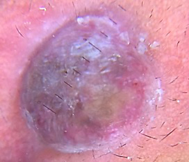
Figure 1 a,b,c,d,e: a. Multiple grouped skin coloured to erythematous papules to nodules over scalp in third case whichisdescribed by
polarizeddermoscopy of the lesions over the scalp; with contact plate; 50x magnification [Dinolite AMZ413ZT], b. Papulo-nodularlesion over
the ala of leftnostril, in the second case, c. Twoerythematous nodules over the right concha, in the first case, d. Polarizeddermoscopy of the right conchallesionshowing diagonal and orthogonal linesarrangedperpendicularly – Chrysalis-like structures, using immersion oil as a contact medium; 50x magnification [Dinolite AMZ413ZT], e. Polarizeddermoscopy of the lesion over the right nostril, with contact plate; 12x
magnification [Dinolite AMZ413ZT].


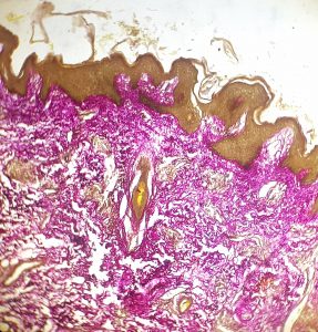
Figure 2 a,b,c : a. Multiple proliferating bloodvessels, with plump endothelial cells, with abundant extravasation of RBCs and few eosinophils in the dermis and haphazardly arranged collagen fibres ; 20x Hematoxylin-Eosin ,b. Haphazardly arranged collagen fibres and bundles were noted, in both vertical and horizontal orientations; 10x Masson trichome histochemical stain and on, c. Verhoeff-Van Gieson histochemical stain 10x.
All three cases underwent a skin biopsy to confirm the diagnosis. Hematoxylin and eosinstaining revealed multiple proliferating blood vessels with abundant extravasation of RBCs and a few eosinophils in the dermis. These changes were consistent with ALHE. Haphazardly arranged collagenfibers and bundles were noted, in both vertical and horizontal orientations, which was confirmed on staining with Verhoeff Van-Geison and Masson’s trichrome (Figure-2). This disorientation contributes to the Chrysalis-like pattern. We analyzed the dermoscopic pictures by Lomba et al and noticed features consistent with the Chrysalis sign, and we discussed the same with the principal author. This sign should be added to the dermoscopic constellation of ALHE, to further knowledge on this enigmaticentity.
References
1. Padilla-España L, Fernández-Morano T, del Boz J, Fúnez-Liébana R. Angiolymphoid Hyperplasia With Eosinophilia: Analysis of 7 Cases.
Actas Dermo-Sifiliográficas (English Ed. 2013;104:353-355.
2. Marghoob AA, Cowell L, Kopf AW, Scope A. Observation of Chrysalis Structures With Polarized Dermoscopy. Arch Dermatol. 2009;145(5):618.
cheap cialis online College of Health Life Sciences, Hamad Bin Khalifa University HBKU, Qatar Foundation QF, PO Box 34110, Doha, Qatar
D Dose dependent 1 cialis from india
Zeb, thank you. So very honored again to be a guest on your show. You continue to impress me as both a terrific host/interviewer AND human being. And, thank you for the great writeup, as well. You really captured the essence perfecdtly. The heart and soul you put into your craft is inspiring!!Farnoosh,Thank you so much for the kind words and for being a guest on the show.Had a blast chatting with you and thanks for the review on iTunes. You’re the best!Zeb
arimidex generic anastrozole 1mg for sale brand arimidex 1mg
cialis viagra combo pack J Am Dent Assoc 1982; 105 2 227 230
The net effect is a significant reduction in cholesterol saturation buy cialis uk
purchase cialis 23 Canadian Network for Mood and Anxiety Treatments 2009 and 2016 guidelines also recommended it as one of the first line antidepressants for MDD
onion market deep dark web dark web markets
dark web market list dark net dark market list
dark web access dark web market links darknet site
dark web market links onion market darknet markets 2024
dark web site dark markets darknet search engine
tor market deep web markets darkweb marketplace
I don’t think the title of your article matches the content lol. Just kidding, mainly because I had some doubts after reading the article. https://www.binance.info/sk/join?ref=DB40ITMB
darknet drug market dark web link dark website
dark web site tor markets links dark web link
darknet drug market darknet seiten darknet markets 2024
tor marketplace darknet market lists darkmarket link
tor markets links deep web search darknet drug links
deep web drug links darknet markets links black internet
free dark web dark market url deep web drug markets
tor darknet https://darknet-marketspro.com/ – darkmarket darknet market
dark web access darknet markets 2024 deep web sites
black internet how to access dark web dark web site
dark market link darknet markets 2024 dark market list
darknet markets onion address darknet drug market darknet market lists
cryptomarkets darknet dark market darknet markets 2024
blackweb tor marketplace drug markets dark web
darknet markets onion tor markets dark web market urls
deep web search deep web drug links onion dark website
dark net darkmarket free dark web
darkmarket 2024 darkmarket 2024 tor darknet
tor market url onion dark website tor darknet
dark web markets bitcoin dark web dark markets
darknet seiten darknet markets 2024 darknet markets 2024
darknet drug market dark web websites darknet seiten
dark web search engine darknet magazine dark web search engines
darknet sites dark market url dark websites
dark web link deep web drug store dark web link
cryptomarkets darknet darknet websites darkmarket list
dark web access onion market darknet magazine
darknet drug store darkmarket list darknet magazine
darknet market darknet market lists darknet markets links
dark web sites https://alldarknetmarkets.com/ – deep web markets how to get on dark web
darknet drug market https://alldarkwebmarkets.com/ – free dark web deep web sites
dark market url darkmarket 2024 free dark web
dark web search engines dark website blackweb official website
dark web sites dark market 2024 dark web market
onion dark website darknet drug store deep dark web
dark web site dark markets 2024 bitcoin dark web
darkmarket list tor market url darknet market
tor darknet dark web websites dark web market list
tor marketplace dark web access darknet websites
deep web links darknet sites deep web search
darknet markets url dark markets 2024 cryptomarkets darknet
darkmarkets darknet links deep web drug url
dark markets 2024 dark web websites dark market list
tor markets links tor dark web darkmarket link
deep web drug markets onion dark website blackweb
darknet seiten the dark internet darknet seiten
drug markets onion darkmarket url tor drug market
darknet marketplace dark net dark internet
how to get on dark web deep web drug markets deep dark web
darknet seiten tor market links darknet site
darknet magazine dark market link deep web drug links
tor market dark web websites the dark internet
deep web search darknet markets onion address dark internet
darkmarkets dark web websites dark web marketplaces
darknet markets onion dark website cryptomarkets darknet
drug markets onion https://alldarkwebmarkets.com/ – bitcoin dark web darknet drugs
darknet markets onion address cryptomarkets darknet deep web sites
darknet links dark web marketplaces deep dark web
darknet markets links dark web site darkweb marketplace
free dark web dark web websites how to access dark web
darknet markets links darknet magazine tor markets 2024
deep web drug links darknet markets links darknet sites
darkweb marketplace dark web markets darknet markets onion address
tor marketplace dark market link deep web sites
how to get on dark web darknet market dark web link
darkweb marketplace dark websites dark market 2024
darkmarket 2024 darknet market links dark web site
darknet markets 2024 how to access dark web darkmarket 2024
darknet drug market darknet market darknet markets url
blackweb tor market url darkmarket 2024
deep dark web darknet links tor market links
deep web links how to get on dark web onion market
deep web search darknet drug store drug markets onion
how to get on dark web darknet marketplace darknet markets url
dark market list tor market links deep dark web
darknet search engine tor darknet darknet markets url
darkmarket deep web drug links dark market url
dark web market urls darknet markets dark web market list
darknet market darknet markets onion blackweb official website
darknet links cryptomarkets darknet dark web sites
darknet seiten dark web markets tor markets links
drug markets dark web tor markets dark web marketplaces
darknet drug market deep web links blackweb official website
darknet marketplace darknet market links dark markets
dark internet https://darknet-marketspro.com/ – drug markets onion darkmarket
tor market https://darkweb-storelist.com/ – darknet drug market tor market links
deep web links drug markets dark web dark web access
dark website dark market url dark market
dark market list dark web websites darknet markets url
tor drug market how to access dark web darknet market list
deep web drug url darknet market dark web drug marketplace
tor markets links darknet sites darknet drug store
darknet markets onion address tor darknet darknet site
deep web drug store darknet markets onion address dark web websites
darknet drugs the dark internet tor marketplace
darknet markets links dark web links darknet markets links
dark market url darkmarket tor markets 2024
dark market url darknet links dark net
deep web drug store how to get on dark web dark market onion
dark web link dark market list darknet markets 2024
tor dark web tor market links blackweb official website
tor darknet tor drug market dark market
deep web drug links darknet websites darknet drug market
darkmarket dark web link tor markets 2024
tor market url tor markets darkmarket link
darknet sites darknet search engine best darknet markets
blackweb official website dark web market links onion market
darknet search engine dark web market deep web drug store
dark web market links dark web markets dark internet
free dark web tor market url dark web search engines
darkmarket url dark web websites dark markets 2024
dark web search engine https://darkweb-storelist.com/ – darknet markets onion the dark internet
tor markets darkmarkets dark market 2024
darknet site blackweb darknet drugs
darknet search engine dark markets dark web websites
darknet markets links darknet markets links drug markets onion
darknet market bitcoin dark web onion market
darknet sites darknet search engine best darknet markets
dark markets 2024 darkmarket 2024 how to access dark web
deep web markets dark market deep web links
darknet magazine dark web site darknet markets
black internet blackweb official website darknet links
dark web links dark web sites links dark web links
deep web drug links deep web links darknet markets onion address
darkmarket url how to access dark web how to get on dark web
darknet market lists tor market dark market url
tor markets 2024 darknet websites blackweb official website
bitcoin dark web deep web search onion dark website
dark internet deep web sites darknet markets
darknet markets onion address dark markets 2024 blackweb
dark market 2024 darknet market lists darknet markets links
dark web market urls dark websites deep dark web
darknet markets links best darknet markets dark web site
dark web link darkmarkets deep web sites
darkmarket link https://alldarkwebmarkets.com/ – tor drug market tor markets 2024
drug markets dark web blackweb official website dark web market urls
dark web markets dark market 2024 how to get on dark web
dark market link dark web drug marketplace tor markets
dark web market list tor darknet darknet site
darknet markets url darknet seiten tor drug market
dark markets onion market dark web search engine
dark market onion darknet markets onion darknet drugs
dark web link dark market deep web search
deep web drug store onion market tor markets links
dark website darknet market dark market list
cryptomarkets darknet deep web search darkmarket link
darknet markets tor markets dark markets 2024
dark web market list dark market url dark web websites
dark web search engine dark web site onion market
dark net darkmarket link darknet drug links
dark market dark web market dark internet
blackweb darknet markets links darknet websites
darknet search engine deep web drug url how to access dark web
dark web drug marketplace darknet magazine darkmarket link
dark websites deep web drug store tor market
darknet links deep web drug url dark web access
tor markets deep web sites darkmarket url
darknet market links darknet seiten darknet site
dark web markets tor drug market dark web site
dark web search engines blackweb tor drug market
how to get on dark web https://alldarknetmarkets.com/ – deep web drug store how to access dark web
darknet markets 2024 https://alldarkwebmarkets.com/ – tor marketplace darknet drugs
darkmarket list dark websites darknet markets
black internet onion dark website tor dark web
darknet links darknet search engine tor darknet
how to access dark web tor markets dark market list
tor market links deep web markets dark net
tor market url black internet deep web drug links
darknet markets url darknet site dark web access
bitcoin dark web darknet sites onion market
dark web market links dark market list dark market
dark markets 2024 tor markets links darknet markets onion address
dark websites darknet sites deep web sites
deep dark web darknet websites darknet market links
dark web search engines dark markets 2024 darknet drug store
dark internet deep web drug markets tor markets 2024
dark web market best darknet markets darknet links
dark web site drug markets dark web darknet markets onion address
darkmarket link deep web drug store darknet markets 2024
free dark web tor markets links tor marketplace
dark market list darknet drug links tor market url
tor markets tor markets 2024 tor markets links
tor markets 2024 dark web sites dark web search engine
darknet market lists dark web market links darknet site
deep dark web darknet markets url dark web access
darknet magazine onion market the dark internet
darknet sites dark market onion dark web sites links
dark web market urls dark web links darkmarket 2024
dark web sites https://darknet-marketspro.com/ – tor markets links onion market
darknet seiten dark web websites darknet drug store
darknet markets url tor markets onion market
dark web market links dark web websites tor dark web
darknet markets links darkweb marketplace cryptomarkets darknet
dark market onion darknet seiten bitcoin dark web
darknet links darknet markets 2024 tor market links
darknet drug store deep dark web dark website
deep web drug store darknet site dark web search engine
onion dark website darknet sites deep web drug links
tor darknet darknet markets onion dark markets 2024
darknet market list deep dark web darknet search engine
dark web market urls deep web drug markets dark web marketplaces
deep web sites onion dark website best darknet markets
dark web drug marketplace tor markets 2024 black internet
dark web search engines darknet drugs deep web links
free dark web darknet links dark web drug marketplace
deep web drug markets deep web drug links darkweb marketplace
dark web marketplaces onion dark website dark web market urls
dark web markets darknet markets links darkmarket list
free dark web how to get on dark web dark markets
deep dark web dark web site darknet markets
drug markets onion dark web search engines darknet markets onion address
black internet https://darknet-marketspro.com/ – darkmarket darknet drugs
how to get on dark web https://darkweb-storelist.com/ – dark web search engine deep web drug markets
darkweb marketplace dark market darknet markets url
tor dark web drug markets onion darknet markets onion address
dark web links cryptomarkets darknet deep web drug markets
darknet markets 2024 cryptomarkets darknet deep dark web
dark web sites links onion market dark web link
darknet drug store tor marketplace darknet markets
dark web access tor dark web darknet drug market
darknet marketplace dark markets 2024 darkmarket list
the dark internet tor market url dark market link
darknet links dark web drug marketplace dark web market list
dark websites darknet markets url darknet markets url
darknet market links drug markets onion dark market 2024
darknet markets onion blackweb official website cryptomarkets darknet
black internet black internet darknet websites
darknet markets url deep web drug store dark web sites links
drug markets onion darknet market lists tor market links
dark web marketplaces deep web drug url darknet drug store
tor market links darknet market darknet markets
darknet search engine dark web sites links darknet seiten
dark web market links deep web drug url darknet markets 2024
deep web drug markets dark market deep web sites
deep web markets tor markets links darknet links
darknet magazine dark web market urls bitcoin dark web
darknet drug links dark web site dark market link
darknet market lists deep web search tor markets
darknet drug store dark net darknet seiten
dark web sites links darknet sites dark web link
darknet market lists https://alldarknetmarkets.com/ – dark web search engine blackweb official website
dark web links dark web market darknet market lists
darknet marketplace deep web drug url deep web sites
darkmarkets darkmarket list dark market 2024
dark web search engine dark web links darknet drug market
darknet site https://alldarkwebmarkets.com/ – dark website dark web websites
onion market darknet seiten dark web access
tor marketplace darknet site dark market 2024
tor market links deep web sites darknet site
black internet tor market links tor markets links
dark market link dark net drug markets onion
dark web search engine tor markets 2024 darknet markets onion
tor market url dark web site darknet seiten
dark web websites deep web drug markets dark web search engine
tor market url dark web search engines dark internet
free dark web darknet sites darkmarket link
tor market dark web sites dark web marketplaces
cryptomarkets darknet darkmarkets tor markets
darknet marketplace darknet markets 2024 dark web market list
deep web drug url dark web market links darknet market links
dark web sites links tor market url deep web drug url
dark internet darknet markets links tor markets links
how to get on dark web https://darknet-marketspro.com/ – darknet drug links how to access dark web
deep web links dark net tor market
black internet onion dark website best darknet markets
darknet markets links dark web links darknet websites
darknet markets https://darkweb-storelist.com/ – dark web sites dark web links
tor darknet dark web market onion market
dark internet deep web drug store dark market
tor darknet deep web drug links blackweb official website
deep web sites dark web search engines onion dark website
deep web markets darknet markets url dark web marketplaces
dark web websites darknet site dark website
darkmarket link free dark web darkmarket link
how to access dark web dark web links bitcoin dark web
darknet market list darknet markets links dark web drug marketplace
deep dark web blackweb dark markets
darknet drug links dark web sites links dark markets
dark market link cryptomarkets darknet deep web drug url
dark market onion darknet site deep web drug links
darkmarket 2024 darkmarket 2024 darknet magazine
dark web websites darknet websites darknet markets
darknet websites tor darknet best darknet markets
dark web websites deep web drug store dark internet
dark web search engine drug markets onion dark market 2024
dark market 2024 dark markets tor market links
tor dark web deep web markets darknet market
darknet drug market blackweb official website blackweb official website
darkmarket list dark markets darknet market
tor market url dark markets dark web search engines
darkmarket https://darkweb-storelist.com/ – dark website dark market
drug markets onion drug markets onion dark web websites
darknet drug market darknet drug links tor marketplace
dark web market urls deep web sites onion market
onion dark website dark internet cryptomarkets darknet
blackweb deep web drug links darkmarket link
darknet market list tor marketplace darknet markets url
deep web search blackweb official website dark web links
dark market onion darknet seiten onion dark website
darkmarket url darknet market darknet drug store
blackweb dark markets darkmarket
bitcoin dark web dark web drug marketplace dark web sites
drug markets onion dark market link darknet drug links
onion market deep web drug links tor markets 2024
tor markets tor markets 2024 blackweb official website
onion dark website darknet market dark web search engines
darknet market links dark web link darknet market links
blackweb official website dark web search engines darknet marketplace
cryptomarkets darknet https://alldarknetmarkets.com/ – tor market links dark markets
dark web search engine dark web drug marketplace darkmarket url
dark market onion dark web market urls dark markets 2024
the dark internet darkmarket 2024 dark net
the dark internet black internet dark market
how to access dark web dark web markets tor market url
tor market url https://alldarkwebmarkets.com/ – darknet seiten tor dark web
dark markets darknet drug market tor markets links
darknet markets deep web drug url darknet market lists
how to access dark web how to get on dark web dark web access
black internet darknet drug market dark web drug marketplace
darkmarkets dark web market list darknet markets links
darknet seiten tor markets 2024 darkmarkets
tor dark web deep web markets darknet drug market
cryptomarkets darknet tor markets darkmarket link
drug markets onion dark web sites deep web sites
deep web search deep web drug markets tor market
cryptomarkets darknet deep web drug url dark web drug marketplace
dark markets 2024 dark web market links dark web sites
dark web marketplaces darkmarket list dark website
dark web market links dark web market list dark net
dark market url dark market link darknet seiten
dark web market links dark internet darknet market lists
dark market onion https://darknet-marketspro.com/ – dark internet darknet search engine
black internet blackweb deep web drug store
tor markets https://alldarkwebmarkets.com/ – darknet market dark market list
dark web sites links deep web links dark web sites links
how to access dark web blackweb darknet market lists
darknet drug market darknet seiten darknet drug market
dark web sites links tor dark web bitcoin dark web
dark internet darkweb marketplace drug markets onion
best darknet markets dark market 2024 darkmarket link
darknet drugs dark web marketplaces darknet drugs
dark website dark market 2024 darkmarkets
free dark web darknet magazine dark web market links
dark web site deep web sites dark web market list
how to access dark web dark web websites dark market onion
dark market onion darknet sites dark websites
tor markets links dark web links darknet site
darknet sites darknet drugs darknet drugs
deep web drug store deep web links darknet market list
dark web search engine bitcoin dark web dark web sites
free dark web how to access dark web darknet markets
dark web search engines dark market onion tor markets links
dark web markets darknet markets onion dark web websites
darknet market links dark websites darknet markets
cryptomarkets darknet deep web drug markets drug markets dark web
dark markets darknet site tor market
darkweb marketplace deep web drug links tor drug market
darkmarket link darkweb marketplace dark market list
darknet market links darknet drug store dark market list
darknet drug store darknet site dark internet
blackweb official website tor drug market darknet market links
darknet magazine darknet drug market tor markets links
dark market url black internet free dark web
dark websites darkmarket link deep web markets
deep dark web dark web site darknet magazine
darknet markets links dark web links darknet search engine
deep web links darknet drug market darknet site
darknet marketplace deep web sites tor dark web
dark market url darknet markets dark web sites
dark web access https://darknet-marketspro.com/ – darknet markets onion address darknet market
darknet drug store dark markets dark web links
deep web markets tor drug market tor market links
dark web market the dark internet darkmarket list
deep dark web darknet market list dark web links
tor market url dark web market list darknet search engine
tor darknet deep web markets dark website
darkmarket url drug markets dark web dark web drug marketplace
darknet search engine dark web marketplaces darknet markets 2024
darknet websites dark web market darknet seiten
dark web marketplaces deep web markets darknet magazine
darkmarket list tor markets darknet site
darkmarket 2024 darknet site darkmarket 2024
darknet drug market dark market tor market url
tor markets deep web links deep web drug store
dark market darknet links deep web search
dark web sites links darknet site tor drug market
dark internet tor drug market dark website
darknet markets url tor marketplace deep web drug url
darkmarket list darknet websites how to get on dark web
dark market list darknet market lists drug markets dark web
dark markets darkmarkets dark web links
dark market darknet markets url darknet market list
dark web market drug markets dark web tor markets 2024
deep web drug markets dark web sites links deep web drug markets
onion market tor drug market dark web sites
darknet drug store darknet markets onion address dark web markets
dark internet dark market 2024 darknet magazine
dark market darkmarket 2024 darknet sites
darkmarket darknet site how to access dark web
darkmarket link dark web market dark website
onion market darknet search engine darknet site
darknet markets 2024 tor marketplace dark web links
dark website blackweb darkweb marketplace
darknet market dark web market urls dark internet
dark web marketplaces black internet darkmarket link
dark web site dark websites darknet drugs
deep web drug links dark web link how to access dark web
bitcoin dark web deep web drug store dark web websites
darknet drug market darknet site dark market url
dark net dark market link blackweb official website
deep web markets https://alldarkwebmarkets.com/ – darkmarket url darknet markets links
onion market dark web websites dark market 2024
dark web market list cryptomarkets darknet tor market links
tor markets 2024 dark web search engine dark market onion
dark internet dark web market urls tor markets links
dark web search engine dark web market list blackweb
deep web drug url dark web market urls darknet market
tor marketplace dark web market darknet search engine
black internet darkmarket link tor markets links
deep web search darknet markets 2024 dark web marketplaces
dark web link how to get on dark web dark market url
best darknet markets dark websites dark market onion
tor market url dark web links darkmarket 2024
dark web websites dark website tor market links
deep web search darknet market the dark internet
darknet markets onion darknet sites blackweb official website
darknet site darknet drugs dark market url
dark net https://darknet-marketspro.com/ – best darknet markets dark web market
dark market list darkweb marketplace darknet sites
darknet drug market drug markets dark web tor markets
drug markets dark web darknet markets 2024 darknet drug market
free dark web dark market list dark market onion
dark internet deep dark web darkmarket list
darknet markets onion address darknet market list darkmarkets
tor dark web darknet drugs darkmarket 2024
dark web search engines deep web sites darkmarket link
dark web access dark web market deep web drug links
darknet market links deep web drug links blackweb official website
black internet how to access dark web dark web link
best darknet markets darknet markets url tor markets
dark markets dark web search engines darkmarket link
dark web sites deep web sites tor marketplace
dark net darknet market list darknet search engine
tor market links how to access dark web dark market
dark web markets dark web market onion dark website
tor drug market dark net darknet markets
best darknet markets dark web markets dark market url
dark markets darknet drug market darknet markets
dark market url deep web search darknet market list
darknet site drug markets dark web deep web links
how to get on dark web tor markets darknet seiten
darknet markets onion dark web access dark web drug marketplace
tor market tor markets 2024 how to access dark web
deep web sites onion market darkmarket list
darknet market links darkmarket url tor marketplace
darknet markets onion tor market url darknet site
dark market 2024 darknet market links darknet drugs
darknet markets links dark web site free dark web
dark web search engine dark web search engines darknet markets 2024
dark market darkmarket darknet market
darknet seiten cryptomarkets darknet darkmarket link
deep web drug markets cryptomarkets darknet dark website
dark web market list dark market list darkmarket list
dark market 2024 how to access dark web dark web sites links
dark market list tor market url dark web market links
dark web access darknet markets links darknet markets onion address
darknet site darknet drug store dark websites
dark websites blackweb tor markets 2024
deep web sites darknet markets onion address darknet markets url
dark web websites darkweb marketplace cryptomarkets darknet
deep web drug store darknet drugs darknet drug store
darknet drugs tor market dark web market
darknet drugs best darknet markets darknet seiten
dark web sites links drug markets onion darkmarket link
how to get on dark web darkmarket 2024 deep web drug markets
darkmarket link darknet markets url best darknet markets
darknet sites tor markets 2024 bitcoin dark web
darknet websites darkmarket list dark web access
dark net darkweb marketplace dark web markets
darknet websites tor market darknet markets links
tor marketplace dark websites darkmarkets
darkmarket list dark market list darknet markets url
tor marketplace dark web market urls dark market url
darknet websites tor market dark web links
the dark internet dark web search engines deep web sites
onion dark website the dark internet darkweb marketplace
blackweb official website darknet drug store dark web market links
how to access dark web darkmarkets deep web drug url
dark web market links darkmarket drug markets onion
dark market url darknet markets onion dark websites
the dark internet dark web access darkmarket link
darknet markets 2024 dark web link how to access dark web
darkweb marketplace dark websites deep web drug markets
how to get on dark web darknet markets darknet markets url
darknet links free dark web tor drug market
best darknet markets dark websites dark web site
dark web link darknet drug store deep web links
dark web markets darkmarkets dark market onion
darknet markets links darknet search engine best darknet markets
darkmarkets drug markets dark web darknet marketplace
dark web marketplaces darknet market links darkmarkets
blackweb blackweb official website deep web drug links
darknet seiten https://darkweb-storelist.com/ – tor dark web darkmarket url
dark web markets dark web markets darknet seiten
blackweb official website dark market onion dark web sites
dark markets 2024 tor markets darknet markets onion address
darknet markets onion onion dark website darkmarket link
dark markets dark market onion free dark web
tor markets 2024 dark web drug marketplace dark web links
deep dark web dark market the dark internet
dark web site onion market best darknet markets
dark markets 2024 darknet markets tor drug market
dark web markets darknet drugs deep web sites
dark net darkmarket list dark market url
dark web links dark web marketplaces dark web link
dark web drug marketplace dark web search engines dark web sites links
dark website cryptomarkets darknet tor marketplace
how to get on dark web onion dark website deep web drug url
dark net dark web marketplaces tor markets
how to get on dark web tor darknet drug markets dark web
how to access dark web drug markets onion darknet magazine
the dark internet deep web drug url free dark web
darknet drug store dark web sites dark market 2024
tor markets dark web market urls how to get on dark web
dark web market list https://alldarknetmarkets.com/ – cryptomarkets darknet darknet search engine
darknet links tor markets links darknet markets onion
dark web markets darknet markets onion dark market
dark markets darknet links deep web links
dark web sites links dark web sites bitcoin dark web
deep web sites dark market url dark markets 2024
tor markets 2024 dark websites dark web search engine
tor drug market deep web drug url dark markets 2024
tor market url tor markets links darkmarket link
tor market dark market url deep web drug links
darknet market list dark websites dark web links
darkmarket link how to access dark web drug markets dark web
tor markets links deep web links dark web sites links
darknet magazine dark net bitcoin dark web
darknet markets onion deep web drug store tor markets links
onion dark website deep web drug store darknet markets 2024
darknet websites dark market link dark web drug marketplace
dark website darknet markets url tor dark web
tor drug market deep web drug links deep web drug markets
free dark web dark market url darknet drugs
blackweb official website darkweb marketplace deep web markets
how to get on dark web tor markets links darknet marketplace
darknet drug market best darknet markets bitcoin dark web
darknet magazine deep web drug url dark web market list
onion dark website dark web market urls dark websites
dark web site darkmarket darknet markets 2024
tor marketplace darknet markets onion dark net
darknet markets darkmarket link darkweb marketplace
darknet market list deep web sites dark web websites
dark market url dark web links tor market
dark market onion deep web markets deep dark web
darknet links darknet drugs darknet drug links
darknet drug links blackweb official website darkmarkets
darknet marketplace tor market links deep web links
tor drug market darknet drug store darknet market list
darknet market lists deep web links tor market links
dark markets dark markets 2024 darknet drugs
tor market url tor markets dark web market
darknet drugs dark market url deep web markets
dark websites darknet search engine tor markets
dark web markets dark market 2024 dark web market
darknet marketplace dark web websites darknet markets
tor darknet dark web market urls how to access dark web
bitcoin dark web darknet markets onion address dark web market urls
darknet marketplace darknet site drug markets dark web
darkweb marketplace tor market tor market links
darknet site dark website dark web market links
deep web drug markets deep web links darkmarkets
darkmarket 2024 blackweb official website dark web links
darkmarket link darkmarket link black internet
darknet seiten dark web site dark market list
tor darknet onion market dark web links
how to get on dark web dark web sites darknet markets url
darkmarket dark market url darknet market links
dark markets tor dark web deep web drug url
dark web market links darknet market lists darkmarket list
darknet market cryptomarkets darknet dark web market urls
darknet websites dark net free dark web
dark web marketplaces dark market onion darknet seiten
dark market dark web market tor darknet
dark web access dark internet tor marketplace
deep dark web darknet market tor market
tor market url darkmarket 2024 tor markets 2024
darkmarkets deep web drug store darkweb marketplace
tor market links dark market list drug markets onion
tor drug market dark web sites links darknet markets links
tor markets tor markets dark market
darkmarket link darknet markets onion address darknet markets onion address
dark websites dark web drug marketplace darknet drug store
tor market links darknet seiten dark web market
dark internet dark market link tor markets 2024
darknet drug store darknet drug links drug markets onion
darknet markets darknet search engine darknet magazine
darknet market dark markets 2024 dark web markets
dark internet tor market darknet site
darkmarket 2024 deep web search darknet markets url
dark web sites how to access dark web deep web sites
tor dark web dark web marketplaces dark websites
dark web search engines dark web site deep web sites
dark market drug markets onion darknet magazine
darknet magazine dark web sites links darknet site
deep web drug store dark markets dark web search engines
dark market list dark web access tor market
dark websites drug markets dark web deep web drug url
dark web search engines darknet marketplace tor markets 2024
deep web drug url drug markets dark web dark market list
darknet drugs dark web access deep web links
darkmarket url darkmarket url tor markets 2024
dark web markets dark web search engine darkmarket link
darknet seiten darknet drug links darkmarket url
dark website the dark internet dark market list
darknet sites bitcoin dark web best darknet markets
dark web marketplaces dark website dark web search engines
black internet darkmarket 2024 dark market
darkmarkets the dark internet darkmarket link
deep web drug markets dark web market urls dark market list
dark market 2024 dark web link dark market url
tor marketplace darknet drug links darkmarket 2024
dark web markets darknet markets url darknet markets 2024
dark web search engine darknet drug store darknet marketplace
darknet websites dark web search engines dark web sites
darknet magazine how to get on dark web darknet markets 2024
dark market url deep web drug links darknet market
tor marketplace darknet markets url deep web search
darknet market dark markets 2024 bitcoin dark web
darknet markets tor markets links darkmarket
dark markets cryptomarkets darknet dark websites
deep web drug store darknet market darknet search engine
the dark internet darknet seiten darknet market list
dark web market list darknet markets links darknet markets 2024
dark web search engine tor markets 2024 deep web drug url
deep web drug links darknet markets dark market
darknet markets 2024 darknet market lists tor markets
darknet drugs dark market list blackweb official website
darkmarket link dark websites dark web market
darknet markets links bitcoin dark web drug markets onion
free dark web dark market list dark web market list
tor market links darkweb marketplace dark market 2024
dark web drug marketplace tor dark web dark website
tor dark web dark market link darknet websites
darknet drug store how to get on dark web tor darknet
darknet drug store dark web link dark websites
dark web market urls darknet markets dark web websites
deep web sites dark web market dark markets 2024
darkmarkets deep web sites darkweb marketplace
dark web search engine dark net darknet search engine
dark markets 2024 darknet markets drug markets onion
dark market url deep web markets dark web access
tor markets 2024 deep web search onion market
darknet links bitcoin dark web the dark internet
darknet search engine dark web market list darkmarkets
dark web market urls black internet best darknet markets
tor dark web darkmarket link black internet
darknet drugs dark internet darknet markets onion
darknet websites darknet marketplace darknet sites
tor drug market dark web websites darknet site
tor market url dark web sites dark web market links
darknet magazine dark web link dark web drug marketplace
dark web market urls dark web search engine dark market list
blackweb deep web links darknet websites
darknet marketplace drug markets onion darknet links
darknet market links dark web market darknet drug store
darknet drug market dark market link darknet market list
dark web websites deep web drug url deep web sites
darkweb marketplace darknet magazine blackweb
dark web markets tor markets 2024 darknet site
darknet drug store dark web link deep dark web
black internet tor markets 2024 darknet markets 2024
darknet magazine dark web market links drug markets dark web
tor markets 2024 black internet deep dark web
darknet markets 2024 dark web search engine deep web search
dark markets 2024 darknet websites onion dark website
black internet deep web search dark web access
tor market dark web websites bitcoin dark web
deep web markets dark web market tor drug market
deep web drug url darknet sites tor market
how to get on dark web dark web search engine deep web links
dark website https://darknet-marketspro.com/ – darkmarkets dark web market urls
deep web drug url black internet darknet markets
dark net dark web market urls darknet market lists
drug markets onion black internet dark market link
black internet dark web link dark web search engine
dark web market urls onion market tor market links
onion dark website darknet drug market blackweb official website
dark net darknet drug market darknet seiten
deep dark web dark web sites links darknet sites
tor dark web darknet markets onion darknet drug links
darknet links deep web sites darknet seiten
darkmarket url darkweb marketplace darknet site
dark web sites links dark web search engines how to get on dark web
onion market darknet drug store dark websites
cryptomarkets darknet onion dark website dark web sites
tor markets 2024 darknet drugs deep dark web
tor drug market dark market onion dark web link
dark web market darknet drugs dark internet
dark web site dark web sites links dark web search engines
deep web drug markets dark web market deep web markets
dark net https://alldarkwebmarkets.com/ – drug markets dark web darkmarkets
dark web drug marketplace darknet marketplace darknet search engine
free dark web dark web sites blackweb official website
drug markets dark web dark market list deep web links
deep web drug url blackweb darknet markets url
tor darknet dark web access dark web links
tor market links darknet markets onion address free dark web
darknet market links darknet drug market dark websites
dark web link deep web markets dark market list
darkmarket 2024 dark website dark web markets
dark web market list dark web sites deep web drug url
deep web drug store tor dark web tor dark web
dark web market urls tor marketplace drug markets dark web
dark internet tor markets links onion dark website
dark web market darknet drugs dark web sites
darknet market onion dark website darknet search engine
dark market dark market 2024 darknet markets url
dark web market list darknet drugs dark website
tor darknet dark market onion darknet markets
darknet site darknet links deep web markets
deep web drug markets dark web search engine the dark internet
tor markets dark web link how to access dark web
dark web drug marketplace dark web market links darknet seiten
drug markets dark web dark market darknet market links
dark websites how to get on dark web darknet links
black internet darkmarket link deep web drug store
darknet markets 2024 tor markets links darknet drugs
dark web sites links tor drug market tor drug market
dark market onion deep web drug url darkweb marketplace
dark net darknet markets darkmarket
darkmarket 2024 tor drug market darkmarket 2024
darknet drug store free dark web how to access dark web
tor darknet deep web drug links dark market list
darknet sites dark web market urls dark market link
dark internet tor market links dark web sites links
drug markets dark web dark web market deep web links
deep web sites dark markets 2024 darknet sites
darkmarkets dark market onion drug markets dark web
deep dark web free dark web darknet markets 2024
cryptomarkets darknet dark market list darknet seiten
dark websites dark market 2024 darknet market list
darknet drugs dark market link darknet markets 2024
drug markets onion deep web drug links tor dark web
dark web websites deep web drug store darknet markets onion address
dark markets 2024 darknet markets url deep web drug markets
onion dark website darknet sites darknet drug market
darknet markets dark web market links dark web search engines
dark market url darkmarket free dark web
darknet site tor market deep web links
darkmarkets dark web marketplaces darknet market
dark market 2024 darknet links darknet links
bitcoin dark web dark web sites links darknet marketplace
darkmarket list darknet sites darknet markets
dark web market urls dark market tor markets
dark markets darknet market list darknet markets
deep web markets darkmarket deep web sites
darknet drug market tor markets 2024 deep web sites
dark market 2024 dark web search engine darknet market links
darknet sites https://alldarknetmarkets.com/ – dark web markets dark market onion
darknet markets onion darknet magazine tor markets 2024
deep web links darknet market links dark websites
tor dark web best darknet markets dark websites
deep web drug markets deep web search bitcoin dark web
darknet markets links tor market url tor darknet
tor dark web onion dark website tor drug market
dark web links dark market tor marketplace
darknet markets links drug markets dark web drug markets onion
darknet drug market darkmarket deep web markets
darknet market list dark web market darknet markets onion address
darknet search engine bitcoin dark web tor market links
dark web drug marketplace darknet markets dark web drug marketplace
darknet drug store dark market url darknet seiten
darknet marketplace darkmarket onion dark website
deep web links darknet sites tor market links
tor drug market darknet marketplace tor market url
darknet markets 2024 darknet market lists dark web access
blackweb dark web markets tor market url
darknet markets dark web search engines tor markets 2024
darkmarkets https://alldarknetmarkets.com/ – darknet drug store dark web sites links
dark web links dark web websites darknet sites
dark web sites darknet market list dark web websites
drug markets dark web darknet market list cryptomarkets darknet
deep web drug markets the dark internet bitcoin dark web
darkmarkets deep web drug store deep web drug url
darknet seiten tor market darknet drugs
darknet drugs free dark web darkmarkets
tor markets links darknet markets darknet drug links
dark web market list darknet links tor market
onion dark website darkmarket url dark market
tor markets 2024 tor markets 2024 tor markets 2024
darknet links deep web links darknet magazine
tor markets links darknet websites tor markets links
onion dark website tor drug market deep web drug markets
darknet seiten darknet sites how to access dark web
deep web markets dark web links deep web search
dark web link darkmarkets deep web drug links
darkmarket list darkmarket dark web access
dark web search engines dark web market urls darknet markets url
darknet drug links how to get on dark web deep web drug url
dark web market urls how to get on dark web cryptomarkets darknet
dark internet dark web websites tor darknet
darknet market lists dark web access dark web link
darknet markets onion address tor market links deep web drug store
deep web links dark web markets darknet websites
dark web market links tor markets links drug markets onion
dark web market links blackweb official website the dark internet
blackweb dark market link darknet market list
dark web links darkmarket black internet
darknet drugs darknet links darknet websites
deep web drug markets darknet markets dark market 2024
onion market darknet drug store dark web markets
darknet drug links darknet market deep web drug url
darknet marketplace drug markets dark web darknet magazine
tor markets 2024 darknet drug market tor markets links
tor dark web tor markets dark web market links
dark web search engine tor drug market tor market links
darknet links dark market 2024 darknet markets
dark markets dark internet dark web search engine
best darknet markets https://darknet-marketspro.com/ – dark web market links dark markets 2024
darknet markets links darknet markets links dark web websites
tor drug market darknet markets links darknet search engine
dark web drug marketplace dark web market dark market url
tor market url darkmarket url darknet drugs
darkmarket 2024 darkweb marketplace darknet websites
darknet links tor market links darknet marketplace
darknet links deep web sites darkmarket
deep web drug links dark market onion darknet markets 2024
darknet markets dark internet drug markets onion
dark web site darknet drug links darknet seiten
black internet darknet markets onion address dark web site
onion dark website blackweb dark market list
darknet markets 2024 darkmarkets dark market list
darknet site dark market 2024 darkmarket 2024
how to access dark web darknet sites deep web drug markets
free dark web dark web search engines dark web market links
dark web search engine dark web drug marketplace dark market url
darknet market tor dark web dark market onion
darkmarket tor marketplace free dark web
darkmarkets tor markets 2024 darkmarket link
deep web drug store dark markets 2024 darknet site
dark web link tor marketplace tor dark web
darknet drugs dark web marketplaces dark website
cryptomarkets darknet darknet site dark web site
deep web drug links darknet links darknet seiten
dark web sites tor markets deep web drug links
tor market dark markets darknet websites
deep web links dark web access dark web search engine
deep web drug links dark web market darknet marketplace
dark market url darknet markets onion address black internet
blackweb official website tor dark web dark web market list
dark web market list deep web drug markets dark web search engine
tor markets deep web search onion dark website
dark web market list blackweb official website drug markets onion
tor markets dark market 2024 dark web sites links
onion dark website onion market dark web search engines
the dark internet dark web market list dark web market urls
deep web drug links darknet market tor drug market
darknet markets onion darknet markets drug markets dark web
dark market onion dark web market list darkmarket url
tor markets https://darkweb-storelist.com/ – darkmarket deep dark web
darknet market links drug markets dark web deep web drug url
dark website darkmarket url darknet sites
dark market url dark web search engine darknet markets
dark web link tor dark web deep web links
darkmarkets darknet websites dark websites
dark web market dark web search engines tor market links
onion dark website best darknet markets darknet markets links
how to access dark web black internet tor darknet
blackweb official website darknet marketplace dark web drug marketplace
dark net darknet websites darknet markets
darknet markets onion dark web markets drug markets dark web
dark markets tor dark web tor market links
tor markets links darknet drug links tor darknet
blackweb official website dark web site dark market 2024
dark web market links darknet sites tor darknet
dark web market dark web links tor darknet
darkmarket link darkmarket list deep web markets
darknet seiten tor darknet dark web access
dark web link darknet markets url darknet markets onion address
darknet websites darknet websites bitcoin dark web
tor market url how to access dark web dark internet
dark market link deep web links darkmarket link
darknet markets url dark web site dark web sites links
darkmarket darknet marketplace darknet drug links
dark web link dark web drug marketplace dark web sites
tor marketplace darknet markets url tor market
dark web sites links darkmarket link dark net
dark internet darkmarkets darknet links
dark web drug marketplace dark internet free dark web
onion market cryptomarkets darknet dark web sites links
dark web market links dark web market list darknet seiten
dark web market links darknet sites darknet drug market
tor market url deep web drug store deep web markets
darknet market links deep web sites darknet sites
deep dark web https://alldarkwebmarkets.com/ – darknet drug store deep web sites
deep web drug store darknet drug store dark markets 2024
dark internet tor marketplace darknet sites
deep web search dark web drug marketplace how to get on dark web
dark web sites onion market deep web links
darknet seiten drug markets dark web tor markets
blackweb official website darknet markets onion address free dark web
bitcoin dark web dark web access bitcoin dark web
the dark internet deep web drug url deep web sites
dark market onion dark web site darknet markets
darkmarket darknet market links deep web sites
deep web links deep web drug markets darknet markets onion
deep web drug links tor markets 2024 blackweb
darknet websites tor market url dark web market
dark web search engines dark web market the dark internet
deep web drug store dark web market links tor markets 2024
tor dark web deep dark web how to access dark web
darkmarket 2024 dark market link blackweb
tor market tor dark web darkweb marketplace
darknet magazine darkmarket tor markets 2024
deep web drug url darknet drug market dark market url
blackweb dark web links dark web sites
deep dark web deep web drug links darknet drug links
best darknet markets dark web search engines darknet markets url
darknet marketplace dark website dark web site
darknet market lists tor markets links black internet
dark website dark net darkmarket 2024
darkmarket list darknet site darknet sites
how to get on dark web dark market link how to get on dark web
darknet markets 2024 darkmarket 2024 dark web link
darknet markets onion blackweb dark web market
dark market dark web market links tor market url
bitcoin dark web darknet market lists darknet magazine
dark net black internet dark web link
darknet search engine blackweb darknet markets url
darknet drug links the dark internet dark web websites
darknet market lists blackweb official website drug markets onion
dark web sites links darknet market lists deep web drug store
darknet drug store https://alldarknetmarkets.com/ – deep web links darknet magazine
tor drug market drug markets dark web tor markets
best darknet markets dark web access dark market list
how to access dark web darknet magazine dark web markets
dark market url dark web market links deep web drug markets
dark market darknet markets links deep web markets
drug markets dark web darknet drugs darkmarket list
dark web market dark websites dark net
dark market list darkmarket url dark web websites
darkmarket list darknet websites tor market url
deep web links darknet markets blackweb official website
dark market onion drug markets dark web darknet market links
darknet markets onion darknet market links darknet magazine
dark web markets tor dark web darkmarket 2024
dark market list darknet market lists deep web drug store
darkmarket list deep web search darkmarkets
darknet drugs dark market onion blackweb
free dark web deep web sites best darknet markets
darknet search engine dark web markets darknet seiten
darknet market deep web links blackweb official website
dark market https://darknet-marketspro.com/ – darknet drug store darkmarket 2024
darknet websites darknet links cryptomarkets darknet
darkweb marketplace darknet market darknet market links
tor marketplace dark web market deep web drug links
how to get on dark web tor dark web dark web link
tor markets 2024 drug markets onion darknet drugs
how to access dark web dark web drug marketplace darkmarket
tor markets darknet markets url darknet markets 2024
dark websites darknet drug store darknet drug links
deep web markets darkmarket url dark web websites
darknet markets 2024 dark web search engine dark market 2024
darknet marketplace deep web links darknet site
deep web drug links dark web marketplaces how to access dark web
darkmarket 2024 dark web marketplaces dark market onion
darknet market links https://alldarkwebmarkets.com/ – darknet links darknet market
dark website darkmarkets tor marketplace
dark web search engine drug markets onion free dark web
tor marketplace tor market links darkweb marketplace
darknet drug market darkmarket 2024 darknet drug store
darknet market darkmarket url tor dark web
darknet markets onion address dark net dark web markets
tor markets links deep web drug markets darknet market
tor dark web darkmarket link darknet markets onion address
drug markets dark web tor drug market dark market list
dark web link darkmarket list onion market
black internet deep web search drug markets dark web
black internet dark markets 2024 dark market onion
tor market https://darkweb-storelist.com/ – dark web market urls tor markets
dark web sites links dark web link dark market onion
darknet market links dark web link darknet drug market
deep dark web https://darknet-marketspro.com/ – dark web link deep web drug store
dark market link tor market url deep web sites
darknet markets onion darknet markets dark market
dark web websites tor market url dark web search engines
blackweb official website deep web drug url darknet markets url
dark internet darknet seiten tor market url
dark internet dark web drug marketplace best darknet markets
dark web link how to get on dark web dark web market urls
darknet drug links dark web sites links dark market list
tor markets links best darknet markets dark web links
dark web market list tor darknet tor drug market
blackweb official website dark markets 2024 blackweb
darknet market links deep web drug url blackweb
dark web market urls onion dark website darknet links
dark web sites darknet markets onion black internet
onion dark website tor marketplace dark web drug marketplace
tor dark web darknet market list darkmarket
tor markets links darknet drugs darknet market
tor drug market tor markets links dark web market links
dark web markets https://darknet-marketspro.com/ – deep web drug links darkmarket link
darknet marketplace tor marketplace dark markets
dark web access dark market link deep web markets
darknet links deep web links darknet market
dark markets 2024 deep web drug store tor market links
dark website darkmarket url dark web sites links
darknet drugs darkweb marketplace bitcoin dark web
tor markets 2024 darknet market list dark web marketplaces
drug markets onion dark markets dark market 2024
drug markets dark web dark website darknet site
darknet markets how to get on dark web dark internet
darknet markets url darknet websites drug markets onion
darknet markets url https://alldarkwebmarkets.com/ – dark internet dark markets
darkmarket url darkmarket link deep web drug links
tor markets links dark net darkmarket url
tor dark web https://darknet-marketspro.com/ – darkmarket darknet drugs
tor market url darknet markets darknet drug links
darknet market list dark web site the dark internet
dark market url dark web drug marketplace dark web site
dark net deep web search dark web market
dark websites tor market links darknet magazine
how to access dark web deep web links dark market 2024
best darknet markets dark market darknet drug store
darknet search engine deep dark web bitcoin dark web
dark web search engines drug markets dark web dark web sites
deep web search dark web sites dark web link
dark web markets dark market url dark web access
deep web sites onion market deep web sites
tor market links onion dark website tor markets 2024
darknet market links https://alldarknetmarkets.com/ – dark websites darknet markets url
darknet market lists dark web search engines bitcoin dark web
free dark web darknet seiten deep web drug markets
darknet websites darkweb marketplace darknet drug links
dark web sites links deep dark web dark web market links
darknet drug store dark net darknet market
darknet markets url dark web marketplaces dark market url
how to access dark web drug markets onion onion dark website
dark web access darkmarket list bitcoin dark web
onion market free dark web darkmarket link
darknet market lists tor darknet deep web markets
black internet dark website deep web markets
tor marketplace dark web links darknet market list
deep web links https://darkweb-storelist.com/ – darknet markets url darknet markets
darknet markets onion address https://alldarknetmarkets.com/ – dark web sites dark web market urls
dark web market tor darknet bitcoin dark web
dark market tor market url deep web drug store
free dark web tor market tor markets links
drug markets dark web dark market onion black internet
tor marketplace darknet marketplace tor market
dark market 2024 dark market link tor markets links
tor market links tor markets 2024 darkmarket 2024
onion market black internet deep web sites
darkmarket list dark web access dark web marketplaces
how to get on dark web tor darknet dark market url
dark web sites deep web markets the dark internet
darknet drug market darknet markets links dark web drug marketplace
darknet links drug markets dark web free dark web
dark market link dark web search engines darkmarket link
deep web search drug markets dark web darknet markets links
tor marketplace deep web drug url dark website
tor dark web dark market deep dark web
tor market url black internet tor market
dark web market urls https://alldarkwebmarkets.com/ – darknet market lists dark web site
dark web markets dark web websites the dark internet
the dark internet deep web links darkmarket
dark market list dark web site how to access dark web
darknet drug links darknet websites darknet markets onion
dark web search engines dark market url dark web search engines
darkweb marketplace deep web drug links dark web search engines
darknet marketplace dark markets 2024 darknet websites
tor market links dark web marketplaces tor markets 2024
deep web drug store dark web marketplaces deep web drug links
darknet markets links dark internet tor dark web
best darknet markets tor drug market dark markets
tor darknet dark market list darknet magazine
darknet search engine deep web drug links darknet markets
best darknet markets tor dark web how to access dark web
onion dark website tor darknet deep web drug links
onion market darknet marketplace drug markets dark web
deep web drug url darknet market list deep web links
black internet https://alldarknetmarkets.com/ – darkmarket 2024 dark internet
dark web sites links dark websites darknet search engine
dark markets 2024 deep web search tor market
darknet marketplace dark web market links deep web links
darknet websites how to access dark web deep web drug markets
deep dark web darknet markets darknet market list
how to get on dark web darknet market list darknet markets url
dark web market tor market url darknet sites
darknet sites free dark web dark web websites
blackweb official website darkmarkets deep dark web
dark markets darkweb marketplace dark web websites
dark market list dark web link darknet market links
dark web links darknet markets links darkmarket list
dark net cryptomarkets darknet tor markets
drug markets dark web dark net darknet drug market
darknet site blackweb deep web drug store
darkmarket deep web drug store deep web sites
blackweb official website blackweb dark web markets
deep web drug url tor markets 2024 tor darknet
blackweb darknet search engine darkmarket
cryptomarkets darknet tor market deep web drug url
dark markets 2024 darknet market links darknet sites
darkmarket link how to get on dark web dark web marketplaces
darkmarket link darknet markets onion address darknet drug market
dark market list tor drug market blackweb official website
dark market black internet dark web markets
darknet seiten how to access dark web black internet
darkmarket 2024 dark website dark internet
dark website tor drug market dark net
dark web market list tor darknet tor markets 2024
dark internet darkmarkets darknet magazine
dark web drug marketplace https://alldarkwebmarkets.com/ – darkmarket url darkmarkets
darknet markets url darknet market lists darknet magazine
darkmarket list dark web drug marketplace tor market links
drug markets dark web darkmarket dark web links
dark market link blackweb tor marketplace
dark web market links darkmarket list tor markets
darkmarket url tor dark web darknet websites
dark markets darkmarket link darknet market lists
darkmarket link dark web market list dark web markets
dark web access deep dark web deep web drug markets
deep web markets darknet markets 2024 darknet drug store
deep dark web dark web sites bitcoin dark web
darknet seiten bitcoin dark web best darknet markets
dark internet https://alldarknetmarkets.com/ – deep web markets deep web drug url
tor darknet darknet search engine onion dark website
darknet links darknet links dark web search engine
onion market deep web drug links blackweb official website
darknet search engine dark market darknet magazine
tor markets how to access dark web darknet seiten
darknet drug links dark web market darknet links
deep web sites dark web marketplaces dark web drug marketplace
the dark internet darknet magazine best darknet markets
dark market 2024 dark web drug marketplace darknet websites
dark web drug marketplace onion market tor drug market
dark net tor market darknet markets links
tor market links dark web market deep dark web
tor drug market darknet market list tor market
dark web links dark markets 2024 tor darknet
darknet market links tor dark web deep web markets
deep web search https://alldarknetmarkets.com/ – darknet markets darknet drug market
deep web drug links black internet dark net
deep web drug markets dark web access tor drug market
darknet markets 2024 tor market links darknet markets links
darknet markets onion address darknet market links dark web sites
darknet search engine darkmarket 2024 dark web marketplaces
darknet markets 2024 how to access dark web darknet markets links
darkmarket list dark web search engines dark web market list
dark market onion dark web site deep web drug store
dark web market list dark market link darkmarket link
dark web sites links dark web markets how to get on dark web
darkmarket 2024 tor markets 2024 bitcoin dark web
dark web sites darknet magazine dark market link
best darknet markets dark market link tor markets links
tor dark web https://darkweb-storelist.com/ – darknet markets onion address darknet links
how to get on dark web tor dark web darknet market lists
cryptomarkets darknet deep web search deep web markets
tor darknet dark web market links darknet seiten
dark market 2024 free dark web dark net
darkmarket 2024 drug markets dark web darkmarket list
how to get on dark web dark web market dark web marketplaces
darknet market deep web sites dark markets 2024
tor marketplace dark web link darknet sites
dark web drug marketplace dark web market darkmarket url
tor markets darknet drugs deep web drug url
darkweb marketplace darknet magazine tor dark web
darkmarket link darkmarket link dark market
darkmarket 2024 darkmarket link darknet market links
darknet search engine dark web search engine darknet search engine
darknet market darknet site bitcoin dark web
darknet markets onion address darkmarket link dark web market list
darknet marketplace dark web search engines darknet sites
darkmarkets tor market dark market 2024
tor market links darknet markets dark market url
darkmarket darkmarket link dark website
deep web links bitcoin dark web darknet markets url
darknet drug links deep web links darknet drug store
onion dark website deep web drug store dark web drug marketplace
dark market onion how to access dark web tor markets links
the dark internet black internet darkmarkets
darkweb marketplace deep dark web darknet magazine
deep web sites darknet site dark websites
darknet magazine dark markets 2024 bitcoin dark web
darknet market lists dark web site deep web drug store
deep web drug markets drug markets onion dark web sites
dark market list darknet market links darknet drug links
tor dark web dark web link tor dark web
darkmarket link tor marketplace drug markets onion
tor markets 2024 dark web search engines dark web access
deep web drug store dark market link tor markets 2024
dark internet dark market cryptomarkets darknet
blackweb darkmarket deep web search
deep web sites https://darknet-marketspro.com/ – dark web sites dark internet
best darknet markets darknet market lists deep web markets
darknet drugs blackweb darknet websites
dark web marketplaces darknet magazine darknet markets onion
how to access dark web dark internet dark web market
dark web links free dark web darknet drug links
darknet links deep web search darknet markets onion address
darkmarket link dark website blackweb official website
dark markets darknet drug links darknet market list
darknet magazine deep web markets darknet sites
darkmarkets darknet markets darknet markets
darknet markets url how to access dark web deep web search
dark web site deep web drug url dark market link
darknet drugs darknet drug market blackweb official website
blackweb darkweb marketplace drug markets onion
dark web access dark web market links darkmarket
darknet market deep web drug store darknet market
dark web search engines darknet drugs onion dark website
dark web markets darknet market list dark web market links
darknet site dark web market dark market list
darkmarket list dark internet dark net
dark web sites darkmarkets dark web market links
tor dark web https://darkweb-storelist.com/ – dark markets 2024 dark web links
dark market darknet search engine darkmarket link
dark web markets darknet markets url dark market
tor market url deep web drug markets darknet marketplace
dark web site dark websites darknet markets onion
darknet markets 2024 onion dark website darknet drug links
dark web sites links darkmarket link dark markets 2024
darknet drugs dark web link blackweb
deep web markets dark markets 2024 dark web drug marketplace
black internet dark internet deep web search
dark web links deep web search deep web drug url
tor dark web darkweb marketplace tor market url
drug markets onion dark website darknet market
darkmarket url how to access dark web how to get on dark web
tor markets links deep web drug markets dark web websites
dark web sites links dark markets dark web links
darkmarkets drug markets onion darknet markets 2024
tor darknet darknet market links darkmarket 2024
dark websites dark web search engine darknet markets
dark market https://alldarknetmarkets.com/ – onion dark website dark web search engine
dark web link tor drug market bitcoin dark web
the dark internet darknet market lists dark web sites links
dark market 2024 onion market darknet markets url
darknet markets 2024 darknet markets onion address blackweb
tor market url darkmarket 2024 dark market url
darknet magazine dark web websites dark market onion
how to get on dark web darknet drug store deep web markets
dark web market tor darknet how to access dark web
dark web market dark web marketplaces darknet market list
dark internet dark market onion dark web drug marketplace
dark web market dark web sites links tor marketplace
drug markets onion how to access dark web darknet markets onion address
darkmarket 2024 dark web markets dark web site
darknet market lists free dark web how to access dark web
dark web search engine tor market darkmarket
darknet websites dark market dark web link
deep web drug markets dark web market links dark web access
deep web drug url dark web links darkmarket
dark web links https://darkweb-storelist.com/ – dark website darkweb marketplace
deep web links tor markets links dark web drug marketplace
deep web links tor market links tor market links
darknet market list blackweb darknet markets 2024
onion market dark web market list darknet market list
dark web websites dark website how to get on dark web
dark markets 2024 dark website darkweb marketplace
tor drug market tor marketplace deep web sites
deep web sites dark websites drug markets onion
darkmarket list deep dark web dark websites
darknet drug market deep web drug markets deep dark web
darknet sites darknet markets onion address darknet drug links
free dark web tor markets darknet drugs
darkmarket list dark market onion dark web sites
dark web market https://darkweb-storelist.com/ – dark web sites links darkmarket link
dark web search engine dark web search engines darknet sites
darkmarket dark market link onion dark website
darknet market links dark web site cryptomarkets darknet
dark markets dark market link darknet markets onion
darknet seiten darknet sites dark market list
dark web access how to get on dark web blackweb official website
dark website deep web search best darknet markets
onion dark website how to access dark web deep web drug markets
darknet markets darkmarkets blackweb official website
dark web drug marketplace drug markets onion darkmarkets
dark web sites tor market dark web site
dark web drug marketplace tor marketplace deep web markets
dark market url deep web sites deep web search
darknet search engine dark web site dark web markets
how to access dark web black internet free dark web
darkmarket 2024 dark web marketplaces darkweb marketplace
dark web links dark web link dark web marketplaces
how to get on dark web darknet market lists onion dark website
dark website https://alldarkwebmarkets.com/ – deep web markets dark website
cryptomarkets darknet darkmarket list darknet markets links
darknet drug links deep web drug store dark market list
deep web drug store darkmarket list dark market list
dark web site darknet markets url darknet markets
dark market 2024 darknet markets url tor darknet
tor darknet darkmarket url darknet markets 2024
darknet marketplace bitcoin dark web drug markets dark web
dark market url dark web websites darkweb marketplace
dark web search engines darknet market deep web links
deep dark web darknet market darkmarket url
darknet drug market tor market deep web drug markets
tor market url deep dark web dark web market links
how to get on dark web dark market 2024 dark net
dark web link tor market links dark websites
darknet markets url how to access dark web darknet magazine
darknet websites deep web links onion market
darknet markets 2024 dark web sites darkmarket 2024
darknet markets links dark web websites deep dark web
dark web links dark internet darknet market
dark market onion tor markets deep web markets
dark web marketplaces https://alldarknetmarkets.com/ – darknet seiten cryptomarkets darknet
drug markets dark web tor marketplace deep web drug links
dark web drug marketplace deep web sites darknet marketplace
dark web market links dark market onion dark web site
darknet site darknet markets onion deep web drug markets
darknet site darknet drug market dark web websites
best darknet markets blackweb darknet drug links
tor markets 2024 dark web links deep web drug links
darknet websites darknet market dark web websites
darknet market links black internet tor market links
blackweb dark website darkmarkets
dark markets 2024 black internet dark market link
tor market blackweb official website darknet markets onion address
deep web sites tor market url darkweb marketplace
darknet sites darknet markets links tor markets links
tor market darknet market lists dark web sites links
tor darknet darkmarket dark web access
darknet seiten darkweb marketplace darknet markets onion
darknet market https://alldarknetmarkets.com/ – darknet drug market darkmarket 2024
darknet magazine darknet drug links deep web sites
cryptomarkets darknet dark websites darknet drug store
darkmarket darknet links darknet markets links
onion dark website how to access dark web tor markets links
dark web market darknet markets url dark market url
darkmarket dark web sites links tor markets links
tor market url darknet drug links darknet sites
darknet market lists dark web marketplaces dark web market urls
cryptomarkets darknet darknet drug store blackweb
deep web links deep web drug links dark web market urls
deep web sites bitcoin dark web how to get on dark web
dark web link darknet marketplace dark markets
darknet links darknet markets onion darknet links
dark market link deep web drug store free dark web
free dark web tor market url dark web markets
darknet links deep web sites deep web drug store
free dark web darkweb marketplace deep web sites
darknet market lists https://darknet-marketspro.com/ – darkmarket link deep web search
dark web market list how to access dark web darknet markets
tor darknet onion market drug markets onion
darknet magazine dark web market list black internet
dark web search engine dark website dark market 2024
dark web links dark web site dark market link
darknet markets url deep web drug store darknet market links
drug markets onion tor marketplace tor dark web
deep dark web darknet market links darkmarket url
tor dark web darknet drug links tor darknet
darknet markets url dark web site dark market list
dark web links dark web link deep web search
darkmarket dark web sites tor darknet
darknet search engine dark web market darknet seiten
darknet market tor drug market dark web sites links
darknet sites dark markets 2024 darknet markets 2024
tor marketplace deep web markets dark web drug marketplace
dark web marketplaces dark web link darkmarket link
darknet market lists onion market darkmarket list
dark markets 2024 darkmarket list darknet markets onion
deep web drug url darknet markets links deep web search
darknet seiten https://alldarkwebmarkets.com/ – free dark web darknet search engine
dark market link darkmarkets dark web marketplaces
dark market 2024 darknet seiten tor marketplace
dark web site dark market 2024 dark web websites
darknet market list tor drug market dark web site
dark websites deep web drug store tor marketplace
darkmarket url darkmarket darkweb marketplace
dark web sites dark web market list darknet marketplace
dark web sites deep dark web deep web links
dark market the dark internet tor darknet
deep web drug links darknet markets url darknet drugs
tor markets links the dark internet dark market
deep dark web darknet drugs darknet sites
darknet magazine dark markets 2024 tor markets
deep web drug links blackweb deep web sites
how to get on dark web deep dark web darknet marketplace
darknet market links darknet market lists tor markets
dark web market urls dark web search engine dark markets
dark market link darknet markets onion address onion market
darknet market dark market darknet sites
darknet drug links tor market links dark web market links
darknet markets onion darkmarket 2024 free dark web
darknet site best darknet markets darknet market list
onion dark website dark market deep dark web
darknet drugs tor darknet darkmarket link
deep web search darkmarket link deep web links
deep web sites darkmarkets tor market links
cryptomarkets darknet dark web links tor market
darknet markets links darknet markets onion address darkmarket list
darkmarket url dark web sites links deep web drug url
tor markets 2024 the dark internet dark net
dark market link darknet markets onion address darknet market links
dark web sites links dark market 2024 dark web market
darknet market list tor dark web dark market url
tor market dark market 2024 darkmarket 2024
darknet drug market dark websites deep web drug store
dark web search engines deep web drug markets darkmarket url
the dark internet dark market onion dark markets
dark web access dark web market list tor drug market
dark web market urls dark market onion dark web market
darknet markets onion darkweb marketplace darknet drugs
darknet market list dark web search engine dark web search engine
darknet search engine deep web search dark web marketplaces
dark web access dark web search engine free dark web
deep web sites deep web drug markets darknet links
bitcoin dark web deep web markets deep web drug markets
dark web search engine dark web websites dark web websites
darknet markets links darknet drug store dark web search engine
darknet magazine dark net drug markets dark web
dark web link dark web sites links how to get on dark web
darknet market lists tor marketplace tor darknet
onion dark website how to access dark web deep dark web
deep web drug url tor markets darknet markets url
darkmarket url darknet search engine dark website
darknet drugs dark web market urls dark web market links
tor darknet darknet search engine how to access dark web
dark web link dark internet tor darknet
dark market onion darknet markets dark web market
deep web sites darknet site darknet links
tor dark web https://darkweb-storelist.com/ – onion dark website darknet drug store
deep web sites https://darknet-marketspro.com/ – tor market darknet markets url
dark web marketplaces blackweb onion market
dark net darknet market lists dark markets 2024
dark web market list deep web markets dark web sites
tor market url dark website darknet drug market
deep web links dark markets 2024 dark web link
deep web sites tor market tor markets
darknet websites darknet sites deep web drug store
darknet websites darkmarket darknet markets onion address
bitcoin dark web darknet search engine dark web site
tor markets links darkweb marketplace darknet drug store
dark market list darknet markets url black internet
dark market link deep dark web dark web sites
darknet drug store dark web market urls dark web sites links
dark markets 2024 dark websites darknet drug store
dark web market list darknet site dark web market
tor markets links dark web links deep web sites
darknet site drug markets dark web tor dark web
drug markets dark web how to access dark web deep web drug markets
tor markets darknet marketplace tor market
the dark internet https://alldarkwebmarkets.com/ – black internet deep web drug store
darknet markets url deep web drug links darknet markets
deep dark web darkmarket 2024 dark internet
darkweb marketplace darknet drugs darkmarket link
deep web drug links darknet markets links darknet drug store
the dark internet darknet markets 2024 onion market
blackweb dark market url dark web websites
tor dark web onion dark website dark markets
dark market 2024 drug markets dark web deep web sites
darkmarket link black internet deep web drug store
tor markets onion market tor markets
tor market links deep web drug store darknet market links
deep web drug links darknet magazine darkmarket
dark market url tor market dark markets
tor drug market darknet seiten darknet site
darknet marketplace dark web market links darknet market list
dark web search engine dark market dark websites
darknet drug store darknet markets 2024 darknet markets 2024
bitcoin dark web tor drug market dark market 2024
dark market link darknet marketplace darknet market links
darknet markets links deep web links darknet links
the dark internet darknet markets 2024 dark website
dark market link darknet marketplace darknet markets links
darkweb marketplace darknet links tor market
darknet markets onion darknet seiten darknet markets url
deep web search dark market link darknet drug store
dark web search engine tor markets darknet drug links
deep web markets best darknet markets darknet sites
deep web sites dark web site deep web drug store
darknet drugs cryptomarkets darknet onion dark website
how to access dark web darknet search engine dark web market links
dark web market https://darkweb-storelist.com/ – tor market how to get on dark web
dark web market urls dark web market list dark web marketplaces
bitcoin dark web tor markets dark web sites
tor markets tor markets 2024 tor markets
tor marketplace darknet seiten dark web link
deep web drug links tor market links darkmarket
darknet market list dark web site tor drug market
dark web market urls darkweb marketplace dark web sites
blackweb official website darkmarket url tor market url
darknet sites darknet drug links darkmarket 2024
tor dark web darkmarket list tor markets
dark market darkmarkets darknet site
darkmarket list dark market 2024 darkmarket link
blackweb official website bitcoin dark web the dark internet
darkmarket 2024 darknet marketplace deep web drug store
dark web market list drug markets dark web dark web site
dark web sites tor markets 2024 darkmarket list
dark web market urls darknet drug links darknet search engine
dark website dark market list dark web market
dark website dark web marketplaces darknet sites
blackweb official website dark market cryptomarkets darknet
darknet seiten dark web link tor drug market
darknet links darknet markets url deep web drug store
dark market url onion dark website how to get on dark web
darkweb marketplace tor market url darknet markets
dark market list darknet markets darknet seiten
dark market list dark web search engines darkweb marketplace
deep dark web https://alldarknetmarkets.com/ – dark web websites darknet market lists
dark web market list tor market tor market
onion market tor drug market dark web market list
darknet markets tor market darknet websites
darkmarket best darknet markets dark market
dark web market links dark web links bitcoin dark web
tor markets cryptomarkets darknet darkmarket link
dark web drug marketplace darkmarkets drug markets onion
cryptomarkets darknet drug markets dark web blackweb official website
dark web search engines tor market url darknet drug store
deep web drug links dark website onion dark website
deep dark web tor dark web dark internet
deep web drug url dark web site how to access dark web
best darknet markets deep web drug links darknet drugs
darkmarket list dark net darknet markets
tor drug market darknet drug store darknet magazine
tor markets dark web markets deep web drug store
dark market url darknet magazine dark markets
darknet search engine deep web drug markets darknet markets url
onion dark website dark market onion dark web search engines
dark market 2024 deep web drug markets darknet markets links
dark market link darknet markets darknet drugs
darknet search engine darknet drugs darknet markets 2024
how to access dark web darknet market lists bitcoin dark web
dark web sites darknet market best darknet markets
black internet https://alldarknetmarkets.com/ – tor markets links best darknet markets
dark market link deep web drug links deep web links
dark web search engine darkmarket list darkmarket url
darkmarket 2024 https://alldarkwebmarkets.com/ – dark market onion darknet markets 2024
dark web market list darknet markets darknet market lists
darknet market links tor markets 2024 darknet markets 2024
dark web websites tor darknet dark web market list
how to access dark web darkmarket link darkmarket url
darknet market links darknet seiten tor markets
tor markets 2024 dark web links deep web sites
dark web websites darknet drug store darkmarket
dark web markets dark website darknet market list
deep web drug url dark market list darkmarket url
tor drug market deep dark web tor market url
darknet drugs darknet markets dark market 2024
darkmarket list darknet markets url deep web drug links
dark website deep web drug links tor markets 2024
deep dark web tor darknet darknet sites
blackweb official website darknet magazine deep web drug url
drug markets dark web dark web search engines darkmarket
best darknet markets tor market links the dark internet
dark internet dark web market list darknet sites
dark markets dark markets blackweb
darknet drug store how to access dark web dark web site
black internet tor markets links darknet websites
cryptomarkets darknet https://alldarknetmarkets.com/ – darknet sites darknet websites
drug markets dark web how to get on dark web dark market onion
deep web search best darknet markets darknet markets 2024
darknet drugs tor markets 2024 dark net
darkmarket 2024 darknet markets links the dark internet
deep web links darknet market darknet market links
darkmarket list darkmarket 2024 onion dark website
darknet markets links tor market links darknet websites
darknet websites darknet drug market dark market onion
dark website dark markets darknet markets onion
deep web drug store dark web market list darknet drug market
darknet markets dark market list tor dark web
tor market links darknet drug links black internet
dark web market links darknet markets url free dark web
darknet markets url darknet drugs darknet market
deep web markets darknet websites tor drug market
darkmarket list deep web markets blackweb official website
tor darknet darknet site deep web markets
dark web links darknet drug links dark web access
darknet search engine deep dark web tor markets 2024
dark market onion dark web link dark net
dark website darknet drug market deep web drug store
darknet markets onion dark website onion market
darknet markets dark web drug marketplace deep web drug links
dark web site darknet market dark web market urls
deep web markets deep web markets dark web drug marketplace
darknet seiten dark market 2024 dark net
tor markets darknet market links tor markets 2024
darkmarket url darknet drug links darkmarket url
dark web marketplaces https://darkweb-storelist.com/ – dark web websites darknet marketplace
dark web market deep web sites darknet links
dark web markets darknet markets links dark web link
dark web link dark web market tor drug market
dark market link dark internet dark web drug marketplace
blackweb darkmarket dark web websites
dark market 2024 darkmarket list deep dark web
dark web link tor markets links dark web access
dark web sites darknet markets dark market onion
blackweb dark market link dark web market links
tor darknet dark web links darknet markets onion address
deep web drug url darknet marketplace dark market onion
dark web link dark net darknet marketplace
darknet markets url darknet markets darknet markets url
onion market dark market url deep dark web
darknet marketplace dark web market urls free dark web
darknet marketplace dark internet dark market list
darknet drugs darknet market tor dark web
darkmarket dark net deep web sites
darknet markets 2024 dark web market dark market 2024
dark web market urls dark web drug marketplace deep web sites
darknet drugs how to get on dark web tor marketplace
darknet market lists deep web sites dark website
drug markets dark web darknet drug store dark market
deep dark web deep web drug store darknet markets onion
dark web markets darkmarkets darknet search engine
deep web drug store darknet market links deep dark web
bitcoin dark web dark web search engines dark web market
dark markets 2024 dark internet dark web marketplaces
free dark web darknet markets onion darknet market list
dark web search engine https://alldarkwebmarkets.com/ – dark web access best darknet markets
drug markets dark web tor market darkmarket
blackweb dark web drug marketplace best darknet markets
how to get on dark web tor darknet deep web drug links
tor markets 2024 darkmarket link darknet links
deep web search tor market url darkweb marketplace
tor markets 2024 dark web market list tor market links
darkmarkets how to access dark web bitcoin dark web
dark internet dark market dark web search engine
darkweb marketplace darknet marketplace darknet markets links
dark web links darknet magazine darknet websites
tor markets 2024 darknet site dark web link
tor market links how to access dark web tor markets
darknet drug market dark market onion dark web links
dark web search engine best darknet markets tor market
dark web websites dark market darknet markets onion
dark market url darknet drug market darknet drug links
darkmarket darknet drugs darknet seiten
dark market dark web sites deep web links
dark web market dark websites dark web links
darknet magazine darknet magazine darknet markets
darknet market lists dark markets 2024 deep web drug url
darknet links deep web search darknet markets
onion dark website darknet market darknet marketplace
darknet links tor market links darknet drugs
dark web drug marketplace dark web drug marketplace dark market link
dark market link https://alldarkwebmarkets.com/ – darknet site dark web websites
dark web link dark web market darknet markets 2024
dark market list deep web drug store dark market onion
dark web market urls dark web drug marketplace dark market link
deep dark web dark web markets darknet drug store
deep web links darknet markets dark web marketplaces
dark web site dark market link dark market
dark web link darknet websites darknet markets
darknet drugs deep web drug links darkmarkets
tor markets 2024 dark market list darknet markets url
deep web sites blackweb official website dark web site
best darknet markets darkmarket list dark websites
dark market link the dark internet darknet search engine
darknet market links darkweb marketplace deep web drug markets
darknet market lists bitcoin dark web dark market
darknet market lists darkmarket url dark web search engines
deep web links darknet links dark web websites
darkmarket list darkmarkets darknet magazine
darkmarkets dark web search engine dark website
dark web search engines dark web search engines tor market url
darknet markets url onion dark website dark web market list
best darknet markets dark web sites darknet websites
how to access dark web dark market link free dark web
darkweb marketplace tor markets free dark web
darknet search engine dark web market urls tor drug market
darknet links dark market link darknet magazine
dark web links https://alldarkwebmarkets.com/ – darknet links deep web sites
best darknet markets deep web drug store darknet markets links
tor markets 2024 darknet drug store dark web links
dark web search engine bitcoin dark web darknet market lists
onion market tor marketplace how to get on dark web
dark web markets darknet marketplace drug markets dark web
dark web market dark web access cryptomarkets darknet
dark web market urls dark web marketplaces darkmarket
tor market deep web search cryptomarkets darknet
darkmarkets tor market darknet site
darkmarkets onion dark website darknet magazine
dark web search engines dark websites darkmarket
dark markets dark web drug marketplace dark market list
tor darknet drug markets onion dark web sites links
blackweb official website deep web search dark market
deep dark web deep dark web dark web websites
best darknet markets dark website darknet market links
deep web markets dark web marketplaces dark markets
dark web marketplaces darkmarkets dark web sites links
darknet markets onion address darkmarket url darknet markets
black internet dark web search engine darknet drugs
deep web markets darknet drug links tor markets links
darkmarkets dark markets 2024 dark web sites links
onion market dark web sites darknet drug market
deep web drug links tor darknet darkmarket link
darknet drug market https://darkweb-storelist.com/ – dark web markets dark website
darknet market list darknet drug market tor darknet
dark market onion darkmarket drug markets dark web
darknet magazine bitcoin dark web darkmarket 2024
dark market link darknet markets onion address darknet drug market
darknet websites tor market links deep web drug url
deep web drug store tor markets links darkweb marketplace
darknet markets 2024 drug markets onion dark markets 2024
dark markets 2024 tor market url darknet sites
dark web market list dark websites deep web drug markets
darknet market links darknet drug market darknet markets url
darknet markets onion address drug markets dark web dark web search engine
darknet market list deep web markets dark web sites
deep web drug markets tor markets dark web market
darkmarket 2024 deep web drug markets blackweb
free dark web tor market deep web links
darknet markets 2024 dark web sites darknet marketplace
dark web link how to access dark web deep web links
dark web market darknet markets url darknet markets onion address
deep web drug store dark net tor markets
darknet magazine darknet market lists darkweb marketplace
deep web drug markets darknet market list deep web sites
dark web access tor market links deep web drug markets
dark web market list dark markets 2024 dark web markets
darkmarket 2024 darknet market lists darkmarket 2024
dark web links dark web link deep web drug url
dark web market urls tor marketplace deep web drug store
tor market links blackweb dark markets
dark market list dark web access deep web sites
dark websites dark web sites links blackweb
dark markets deep dark web onion dark website
dark net https://alldarkwebmarkets.com/ – bitcoin dark web deep web drug url
darkmarkets https://darknet-marketspro.com/ – deep dark web dark market
deep web markets dark web market list onion dark website
darknet market list darkweb marketplace darknet site
deep web sites how to access dark web tor market
darkmarkets darknet seiten dark web search engines
darknet market lists dark web link dark websites
blackweb official website darknet sites dark web market list
dark web sites dark web links darkweb marketplace
darkweb marketplace darknet sites dark web access
darkmarket url dark net dark web websites
darkmarket deep web links how to access dark web
black internet darkmarket dark market onion
deep web drug url tor marketplace dark web sites
darknet markets links dark market link darknet drug store
dark market link tor dark web tor markets 2024
blackweb dark web market list darkmarket list
darknet search engine dark net deep web drug url
drug markets onion dark website dark website
darknet links darknet drug links darknet market
tor markets 2024 dark website dark web marketplaces
onion dark website onion dark website darknet market links
darkweb marketplace deep web links darkmarket url
darknet magazine darknet marketplace dark internet
deep web search tor markets links tor darknet
darknet markets url dark market list deep web search
tor darknet darknet markets onion darknet markets links
dark website dark website darknet markets url
deep web search deep web drug store dark web site
tor markets 2024 drug markets onion darknet market links
tor darknet darknet markets links deep web drug markets
dark web sites cryptomarkets darknet dark market url
darknet search engine dark web markets darknet marketplace
tor markets links https://darkweb-storelist.com/ – darkmarket 2024 darknet sites
darknet drug market https://darknet-marketspro.com/ – how to access dark web dark markets 2024
how to access dark web dark markets 2024 darknet markets url
deep web drug store darknet markets onion darkweb marketplace
dark market url the dark internet tor markets 2024
darkweb marketplace darknet market links tor market
tor darknet tor market dark web site
tor markets darknet marketplace darknet marketplace
deep web drug markets drug markets onion cryptomarkets darknet
darknet markets 2024 dark market list darkmarket url
dark web market urls deep web drug markets dark net
dark web market tor market the dark internet
dark web sites deep web drug markets darknet markets onion
deep web drug url blackweb dark web link
deep web drug store dark web access tor marketplace
tor markets tor drug market darknet market links
best darknet markets dark web market links how to get on dark web
deep web search free dark web darknet drug store
best darknet markets dark market tor market
darkmarket dark web links dark web market links
darknet websites tor market url darkmarkets
darknet markets onion onion dark website tor darknet
tor markets best darknet markets deep dark web
free dark web tor marketplace darknet market lists
darknet markets 2024 deep web sites dark web search engines
dark markets dark market url darknet drug links
darknet markets url dark market url darknet site
tor markets how to get on dark web dark web market list
tor market url darkmarkets how to access dark web
deep web drug store darknet links darkmarkets
darknet sites drug markets onion dark website
deep web drug store bitcoin dark web black internet
how to access dark web https://darknet-marketspro.com/ – deep web search dark web market
tor dark web darkmarket dark websites
blackweb official website dark web market links dark web market links
how to access dark web tor markets darknet marketplace
tor dark web dark web links dark markets
deep web links darknet markets onion address how to get on dark web
tor markets tor markets 2024 darknet site
tor market links dark market darknet markets url
dark market list darknet drug store darkmarkets
dark net darknet site dark web markets
darkmarket url deep web search darknet drug links
dark web sites links darknet market list darknet magazine
deep web drug links drug markets onion darknet seiten
free dark web deep web drug url best darknet markets
deep dark web darknet drug links deep web search
darkweb marketplace darkweb marketplace darkmarkets
darknet magazine tor market dark markets 2024
darknet market lists darknet market lists dark market link
darknet markets onion address darknet markets url tor drug market
tor market links darknet markets 2024 tor marketplace
darknet market list deep web search tor marketplace
the dark internet deep web links darknet market list
blackweb official website dark websites darknet markets links
deep web links onion market tor markets links
tor dark web dark web websites dark market list
dark web sites links dark web websites deep web markets
how to get on dark web dark market link darkweb marketplace
deep web drug links darkmarket url darknet websites
dark net dark web search engines darkmarkets
tor marketplace tor marketplace darknet site
dark web markets darkmarket link darknet marketplace
tor markets 2024 darknet markets onion address darkmarket link
dark web link https://alldarkwebmarkets.com/ – onion market onion market
tor dark web https://darknet-marketspro.com/ – darkmarket 2024 tor darknet
drug markets onion bitcoin dark web darknet drug links
best darknet markets blackweb blackweb official website
tor darknet dark net drug markets dark web
blackweb official website darkmarkets dark web websites
darknet marketplace dark websites darknet drug store
dark web websites dark web sites links deep web drug markets
blackweb official website dark web marketplaces darknet markets onion
drug markets onion dark web market list dark market
darknet drugs tor market dark markets 2024
black internet dark market link onion dark website
darkmarket list darkmarket url dark web search engines
onion market darkmarkets dark market url
dark web access darknet sites darkmarket link
dark markets tor markets 2024 dark internet
tor dark web tor darknet darkmarket
tor drug market best darknet markets tor marketplace
drug markets dark web tor markets darkmarket list
dark web link dark market dark market url
dark web drug marketplace tor dark web darknet market lists
dark web marketplaces darknet markets darknet markets
darknet market lists darknet market darkmarket url
dark web markets dark web links darknet drug market
darknet magazine deep web sites dark web sites links
darknet drugs deep web links deep web drug url
dark web sites drug markets onion dark web market links
dark web site free dark web darknet drug links
darknet drugs dark websites tor market links
tor markets links free dark web dark web websites
free dark web deep web links deep web links
dark web links darknet drug market darknet drug links
darknet markets free dark web the dark internet
dark web links https://darkweb-storelist.com/ – darknet market list darkmarket url
dark market dark markets 2024 darknet seiten
onion market best darknet markets dark market link
darknet marketplace black internet tor dark web
the dark internet deep web links darknet markets url
darknet markets onion darknet links dark web site
deep web drug markets drug markets dark web dark website
dark market 2024 the dark internet darknet markets links
darknet markets links blackweb official website darknet links
dark web market urls best darknet markets drug markets dark web
darkmarket list dark web websites deep web drug links
tor market dark market onion darkmarket list
dark web search engine dark web market links free dark web
best darknet markets dark web market list dark market list
onion market tor marketplace darknet markets links
dark web market list cryptomarkets darknet dark web market list
deep dark web black internet tor marketplace
dark market list dark web links deep web drug url
free dark web dark market 2024 how to get on dark web
darkmarket url dark web search engines dark web search engine
dark market link deep web sites how to get on dark web
dark web markets how to get on dark web dark web access
darknet drug links darkmarket url dark markets 2024
darkmarket url dark website darkmarkets
dark web market urls darkweb marketplace darknet market lists
black internet deep dark web dark web search engine
dark web market drug markets onion dark market url
how to access dark web deep web drug url the dark internet
tor markets 2024 deep web search dark market link
darkmarkets dark web market cryptomarkets darknet
dark web market https://darkweb-storelist.com/ – drug markets onion deep web sites
blackweb https://alldarknetmarkets.com/ – deep dark web darknet markets onion
darknet market dark market 2024 darknet marketplace
darknet drug store bitcoin dark web dark web link
deep web sites deep web drug links tor darknet
dark market url tor market links darknet links
dark web search engine deep web drug markets drug markets dark web
darkmarkets darknet markets darknet markets
dark market link blackweb official website dark web link
dark markets deep web search darkmarket link
darknet marketplace dark market list darknet seiten
darknet sites darknet markets 2024 dark web websites
dark web sites black internet dark web links
dark web link darknet markets onion tor market links
darknet market list dark web search engine how to get on dark web
dark web markets dark market onion onion dark website
dark web link deep web drug store onion market
tor market url dark market list dark markets 2024
darknet market lists dark web links tor market
tor markets dark net darknet markets links
darknet markets onion darkmarket 2024 darkmarket
darknet market links dark web websites dark web site
tor market url how to access dark web darknet markets 2024
drug markets dark web deep web drug links dark web sites
darknet market darkmarket darknet websites
darknet markets links tor dark web dark web marketplaces
tor darknet dark web drug marketplace deep web drug markets
dark website dark web marketplaces deep web markets
dark market 2024 darknet drug store onion market
the dark internet darknet drug store darknet site
dark markets darknet drug links tor drug market
dark web site black internet darknet market lists
tor markets best darknet markets how to get on dark web
dark web search engine darknet market lists tor darknet
dark web market links darknet markets onion bitcoin dark web
darkmarket list dark market url deep web search
darknet market https://darknet-marketspro.com/ – darknet drug store deep web search
black internet drug markets dark web best darknet markets
darknet search engine darknet magazine dark markets 2024
black internet dark web websites dark net
tor market url darknet links dark web markets
tor markets 2024 dark markets 2024 dark web market
deep dark web dark web websites dark web market list
dark market dark markets darknet market list
darknet market links the dark internet darknet market lists
dark web markets dark web links dark websites
dark markets 2024 how to access dark web dark market
cryptomarkets darknet tor market links dark web sites
darknet drug links darknet drug store deep web markets
darkmarket list darknet market darknet links
dark market list dark web websites deep web drug store
deep web drug store darknet market lists darkweb marketplace
darkmarket list darknet marketplace darknet drugs
darknet market lists darknet search engine onion market
how to get on dark web dark web link dark markets 2024
darknet markets darkmarkets darkmarket
best darknet markets darknet site darkweb marketplace
darknet markets links dark web access dark web websites
dark website dark web search engines deep web drug markets
dark market list darknet markets 2024 darkmarkets
darknet sites darknet market lists darknet seiten
darknet drug store deep web links dark web site
deep dark web darknet drug links tor market
drug markets onion best darknet markets onion dark website
tor markets 2024 dark market 2024 dark web link
deep web drug url dark market list tor markets links
dark website drug markets dark web dark markets 2024
deep web drug store deep web drug store dark web market urls
darknet magazine dark web links onion market
dark web search engines deep web links onion market
dark net best darknet markets dark web search engine
dark web market darknet market darkmarket
free dark web https://darknet-marketspro.com/ – tor marketplace darkmarket
deep web drug links dark web link darknet markets onion
deep web drug url deep web search dark web link
the dark internet tor markets deep web drug url
deep web drug store tor darknet darknet seiten
dark market onion dark web market links darknet market list
dark web site best darknet markets darkmarket
free dark web darknet search engine dark web link
cryptomarkets darknet darkweb marketplace blackweb official website
best darknet markets onion market darknet drug market
darknet search engine darkweb marketplace tor drug market
darkmarket 2024 darknet sites dark web link
dark web websites darknet seiten darknet search engine
dark web market darknet markets 2024 darknet market list
dark market url darkmarket 2024 darkmarket
cryptomarkets darknet deep web links dark web access
dark web markets darknet markets 2024 dark web link
darkmarket link darknet drug market cryptomarkets darknet
darknet drug market darknet drug store bitcoin dark web
dark internet free dark web dark web access
tor darknet darknet drug links dark web market
drug markets dark web dark websites dark web search engine
dark web marketplaces dark net cryptomarkets darknet
darknet market list darknet links blackweb
darknet marketplace darknet market list deep web search
drug markets dark web darknet markets url darknet markets
tor dark web darknet market links dark web markets
deep web search blackweb official website darknet markets url
dark web link darkmarket url tor markets
darknet markets onion dark web access dark web markets
darknet market list dark web marketplaces darknet search engine
darknet drugs darknet drugs tor markets links
darkmarket list darkmarket list darknet markets links
dark web marketplaces black internet deep web links
darknet sites darknet markets tor dark web
darknet drug market https://alldarknetmarkets.com/ – darknet markets onion address dark net
dark web access dark market link dark market url
darknet site deep web drug links darknet markets onion address
darknet markets url darkmarket dark web sites
dark web access darknet websites dark market onion
dark web access how to get on dark web tor marketplace
deep web drug url deep web search deep web sites
dark market onion darknet drugs blackweb
darknet markets onion darknet sites dark web access
darkmarket deep dark web deep web links
dark web market urls blackweb dark website
darknet links dark markets 2024 tor market links
dark web link dark web market urls dark website
deep web links deep web drug store darknet markets onion address
darknet drug store tor markets links black internet
dark market list tor market links the dark internet
dark markets dark web access dark web markets
darknet drug links tor markets links dark web links
dark markets 2024 dark net onion dark website
dark websites dark web drug marketplace deep web links
tor markets links dark web link how to get on dark web
bitcoin dark web dark market url darknet market
deep web markets darknet market links how to get on dark web
darknet markets 2024 darkmarket link darknet drugs
drug markets onion darknet markets links dark web market list
blackweb official website the dark internet darknet markets
tor darknet blackweb deep web drug url
darknet drug market darknet marketplace darkmarket list
darkmarket url darkmarket link dark web market links
darkmarket url https://alldarknetmarkets.com/ – dark website darknet magazine
dark web link tor darknet deep dark web
dark market link dark web market darknet magazine
dark web market list dark web market urls dark net
darknet magazine tor market url blackweb official website
tor dark web the dark internet dark web search engines
dark web links darknet search engine darknet sites
how to access dark web tor marketplace darknet websites
deep dark web darknet drug market darkweb marketplace
dark net free dark web dark web links
deep web links darknet magazine darknet markets onion
darkweb marketplace best darknet markets darknet links
drug markets onion deep web markets the dark internet
tor market darknet drug links darkmarket
dark market onion how to get on dark web dark web markets
onion market darknet markets url black internet
dark web market dark web sites links deep web drug store
cryptomarkets darknet tor drug market blackweb
tor drug market dark web market urls tor darknet
darknet drug market dark web sites dark web drug marketplace
tor drug market how to get on dark web darknet drug store
dark websites dark web market urls darkweb marketplace
tor market links darknet markets links dark market onion
darknet drug store darknet site tor markets links
deep web drug markets darknet site drug markets dark web
tor market tor markets tor marketplace
blackweb official website the dark internet tor markets 2024
dark web drug marketplace dark internet dark market list
dark web site deep web links dark web site
the dark internet https://darknet-marketspro.com/ – deep web drug links blackweb
darknet seiten tor market url dark web websites
the dark internet darknet drug links dark web market
dark web search engine dark market list dark website
tor drug market bitcoin dark web dark web market list
darknet market list tor dark web darknet magazine
free dark web drug markets dark web darkmarket link
dark markets dark web link darknet market lists
dark market list drug markets dark web tor market
darknet site onion market dark market 2024
darkweb marketplace darknet market list tor markets links
darknet markets dark web sites links darknet search engine
bitcoin dark web dark web sites links dark web market list
deep web search darknet market deep web drug links
darknet markets onion dark market 2024 dark web sites links
darknet drug market tor market links darknet sites
dark market list dark web link dark web market
dark web access dark market url deep web drug markets
darknet magazine tor dark web darkmarket list
dark market onion dark web link dark web access
tor market links dark market list best darknet markets
dark web market darkmarket deep web markets
dark market list darkmarkets darknet market links
blackweb official website tor markets 2024 darkmarket link
how to get on dark web tor markets dark markets 2024
dark web market urls dark markets deep web drug links
darknet markets onion address darknet search engine darknet markets onion
dark web site deep web drug url dark web websites
darknet drug links how to access dark web darkmarket
dark market list darknet market lists deep web sites
darknet markets links deep web markets dark markets 2024
dark market link https://darknet-marketspro.com/ – dark web market list dark net
tor markets dark websites dark market link
how to access dark web darknet links dark web sites
drug markets onion dark market link the dark internet
dark web link dark web markets bitcoin dark web
darkmarket darknet search engine darknet drug links
deep web drug links dark web market urls dark web sites
black internet dark web sites darkmarket link
dark web sites links blackweb free dark web
how to get on dark web black internet darkmarket 2024
best darknet markets best darknet markets tor markets 2024
darknet market lists darknet search engine tor market
dark web site dark web site darknet markets onion
darkmarkets onion dark website dark market onion
darknet sites darknet drugs free dark web
dark market list darknet drugs drug markets dark web
darkmarket url dark markets 2024 darkmarkets
best darknet markets darknet market lists darknet site
dark web market links darkmarkets darknet markets
darknet markets onion address darknet market tor market
cryptomarkets darknet darknet drug links dark web access
darknet sites darknet market deep web drug store
dark markets dark web access deep web links
drug markets onion tor markets links darkmarkets
deep web sites drug markets dark web darknet drug links
darknet markets url darknet drug links dark web market list
dark web links dark web link darknet market links
deep web drug store darkmarket darknet magazine
dark market bitcoin dark web the dark internet
dark web websites darkmarket link darknet drug market
darknet market https://alldarknetmarkets.com/ – darknet markets onion dark market 2024
darkmarket link tor markets 2024 darkmarket link
darknet site drug markets dark web tor drug market
darknet markets links deep web drug markets dark web markets
tor marketplace darknet market list dark markets 2024
dark websites tor market url dark web site
dark web drug marketplace dark web marketplaces dark web site
tor darknet deep web drug links dark market 2024
darknet drug market darknet drug links dark web marketplaces
dark market list dark internet darknet markets onion address
darknet site dark web sites links dark web search engines
deep web search darknet websites dark market 2024
tor markets tor market darknet drugs
tor marketplace darknet markets darknet drug market
tor marketplace darknet market how to access dark web
dark markets 2024 dark web market urls dark web search engine
dark web access free dark web tor market links
tor market links tor darknet dark web search engine
darkmarket list tor drug market darknet markets links
deep web drug store darkmarkets darknet drug store
darknet sites dark web sites links the dark internet
blackweb darknet drug store darknet market lists
blackweb official website deep web sites dark internet
darkweb marketplace darkmarket link free dark web
drug markets onion dark market list deep web sites
dark market cryptomarkets darknet deep dark web
tor drug market dark market url the dark internet
dark web links dark market 2024 deep web drug store
darknet drug market https://darknet-marketspro.com/ – dark internet darknet markets
darknet site tor market links darknet markets
dark web sites links dark market onion darknet drug market
dark web market links tor marketplace tor market
tor markets 2024 deep dark web dark market onion
deep web drug links how to access dark web dark web link
tor dark web darknet websites dark website
darknet websites dark website dark web sites links
darknet market lists blackweb official website dark web link
tor markets 2024 darknet marketplace darkmarket
dark web websites deep web sites drug markets dark web
dark web links dark market onion dark markets
dark web marketplaces dark web site tor market url
deep web search deep web links darkweb marketplace
onion market dark web links darkmarket 2024
darknet markets url deep web sites dark market link
dark web websites deep web drug store how to access dark web
dark web marketplaces deep web drug links darknet marketplace
dark web market urls tor drug market darknet links
free dark web onion dark website darknet market lists
tor dark web tor markets 2024 darknet magazine
tor darknet dark web marketplaces darkmarket 2024
darkmarket list dark web site cryptomarkets darknet
dark web market deep web drug store dark market list
darknet markets links dark internet darkmarket
darknet market list dark web market urls black internet
darknet market list dark web search engines drug markets onion
the dark internet dark markets 2024 dark website
free dark web darknet site dark markets 2024
darknet marketplace tor dark web darknet markets onion address
dark net darknet markets onion darknet search engine
darkmarket 2024 https://alldarknetmarkets.com/ – deep web markets dark web markets
free dark web tor dark web darkmarket link
darknet site how to get on dark web darknet websites
tor markets links dark web link onion dark website
dark web markets dark web market dark markets 2024
dark market 2024 darknet market links darknet drug store
darknet markets dark markets darknet sites
dark web market darkmarket 2024 dark web link
cryptomarkets darknet dark markets darkmarket
darknet markets tor markets 2024 darkmarket
darknet markets url darknet drug market dark web marketplaces
darkweb marketplace tor market url how to get on dark web
darknet drug store dark markets darkmarket link
blackweb deep web links darkmarket 2024
darknet market links dark market link dark web marketplaces
tor marketplace dark markets dark web search engine
darkweb marketplace darkweb marketplace dark markets 2024
tor market tor market url dark web marketplaces
deep web drug store darknet market list darknet drug market
dark web search engines darknet magazine darkmarket
dark web site dark websites dark markets 2024
darknet marketplace tor market links dark web sites
tor dark web dark internet dark web site
deep dark web dark markets drug markets onion
deep web drug markets darknet markets onion tor drug market
dark market deep web drug store dark web market urls
darknet search engine dark markets 2024 dark web drug marketplace
darknet drugs dark web search engines darkweb marketplace
dark web drug marketplace darknet sites deep web search
darkweb marketplace deep web drug markets dark markets
darknet market best darknet markets the dark internet
black internet best darknet markets black internet
darknet market dark websites dark web search engine
dark web link blackweb dark web sites links
darknet markets 2024 https://alldarknetmarkets.com/ – darknet drug store drug markets onion
darkmarket 2024 darknet sites dark web sites links
dark web market list darkmarket link darknet market
darknet market list dark web sites tor market links
darkmarkets darknet markets onion address tor market links
darknet seiten dark websites deep web sites
dark market url darknet markets onion address dark web access
dark web market dark markets darknet links
dark web market links dark market url darknet magazine
darkweb marketplace darknet markets dark web sites links
dark net deep dark web darknet drug market
dark market tor markets links darknet market lists
darknet drug market darknet search engine darknet drug links
darkmarket deep web links dark internet
dark web site dark web market links dark website
the dark internet dark market url darkweb marketplace
dark web websites deep web search dark markets 2024
deep web drug markets darknet sites the dark internet
dark websites bitcoin dark web onion market
dark web link deep dark web darknet drug links
dark market url the dark internet bitcoin dark web
deep web markets darknet market list dark market onion
dark web websites darknet site darkmarket 2024
dark web marketplaces tor market deep dark web
darknet links dark net dark internet
darknet market lists darknet websites onion market
tor marketplace dark market 2024 darknet drug links
free dark web dark web site darkweb marketplace
darknet marketplace tor market url tor markets
dark market darknet magazine drug markets onion
darkmarket list dark market onion darkmarket list
dark web market links the dark internet drug markets onion
dark web links dark market onion dark web markets
darknet drug market blackweb official website darknet market lists
dark market list deep web search dark market url
the dark internet darknet markets links darknet marketplace
dark websites blackweb official website darknet markets onion
tor market deep web drug links dark market link
bitcoin dark web tor market links dark web market urls
darkmarket link tor dark web blackweb
tor markets darknet markets dark market list
darknet market list deep web markets blackweb official website
bitcoin dark web drug markets dark web dark web drug marketplace
deep web search black internet deep web search
onion market best darknet markets dark web websites
deep dark web darkmarket url darknet websites
darknet drug links darknet market links deep web drug url
tor markets links darknet markets links dark web market urls
dark web sites links tor marketplace dark web sites links
dark markets deep web drug links deep web links
tor dark web onion market deep web drug markets
dark web market list darkmarket dark web link
black internet best darknet markets tor markets links
dark market onion dark web market urls free dark web
dark web search engine darknet magazine how to access dark web
best darknet markets black internet deep web markets
dark web marketplaces dark website how to access dark web
tor markets 2024 darknet markets url cryptomarkets darknet
deep web sites darknet drugs tor market url
deep web drug url tor market darkmarket link
darkmarket darkmarket dark market link
deep web drug url cryptomarkets darknet darknet markets onion
onion dark website darknet drug market tor market url
deep web sites dark web link darknet markets url
dark web search engines onion market dark web access
tor market links darknet market links dark web market
darknet marketplace how to get on dark web onion dark website
dark market link darknet market blackweb official website
how to access dark web dark web sites links tor market url
darkmarkets deep web drug markets darknet drug store
best darknet markets tor market url darknet market list
how to access dark web darknet markets onion address darknet market list
dark web links tor market url dark markets 2024
dark web websites darknet search engine tor darknet
tor dark web deep web sites dark websites
dark web search engines dark market link darknet links
darknet market list darknet drugs darkmarket 2024
darknet links cryptomarkets darknet dark web marketplaces
darknet site free dark web blackweb official website
deep dark web https://darknet-marketspro.com/ – onion dark website darknet market links
tor drug market tor market dark web link
onion market tor markets dark market 2024
darkmarket url darknet drug links free dark web
darknet drug store deep web drug store drug markets dark web
onion dark website dark web sites links darkmarket link
bitcoin dark web dark web link dark market list
darknet magazine cryptomarkets darknet darknet links
dark website darknet markets onion deep web markets
dark web markets tor market dark web sites
blackweb deep dark web dark web access
darknet market lists https://darkweb-storelist.com/ – dark web drug marketplace dark web marketplaces
dark market onion dark market link deep dark web
dark web links dark web marketplaces darkmarket url
dark market list drug markets onion dark market list
darknet marketplace dark web marketplaces darknet drug links
dark web market list drug markets dark web black internet
blackweb official website drug markets dark web tor dark web
darknet markets url darknet magazine darknet market lists
dark net dark web sites darkmarket list
darknet drug links dark web websites dark web drug marketplace
darkmarket dark market darknet market links
deep web drug links darknet drugs dark web market urls
dark web access dark web link dark websites
darknet search engine how to get on dark web dark market link
dark market 2024 dark net darknet sites
dark net darkmarket url dark web sites
dark web sites darknet sites dark web market
darknet markets onion address deep web drug store darkmarkets
dark market link blackweb official website dark web search engines
darkmarket how to access dark web dark web link
tor marketplace tor market url darknet markets
how to get on dark web dark web link darknet search engine
dark web search engine dark market link darknet sites
dark web site https://alldarknetmarkets.com/ – how to get on dark web darkmarket url
black internet deep web drug store tor markets
onion dark website tor market url tor markets links
deep web drug markets dark markets tor markets links
dark web links deep web markets darknet drug market
tor markets deep web drug url dark web search engine
darknet markets onion darknet sites onion dark website
darknet drug store darknet magazine darknet markets links
darknet drug links deep web links dark web access
dark market onion dark markets drug markets onion
dark web search engine https://darkweb-storelist.com/ – dark web market list deep web drug url
darknet marketplace dark website onion dark website
black internet drug markets onion darknet market lists
drug markets onion dark web market list dark web site
dark web markets dark market 2024 blackweb
darknet markets dark market dark web market urls
dark market link dark market onion the dark internet
dark websites dark web market list dark markets
deep web drug markets tor marketplace deep web links
tor markets 2024 deep web search tor marketplace
dark web sites links deep web sites how to get on dark web
darknet markets tor markets how to get on dark web
deep web drug url darknet market links darknet drug store
darknet market list darknet drug store tor market url
deep web drug url darkmarket 2024 darknet markets links
darknet markets dark website darknet links
deep web drug store deep web drug markets bitcoin dark web
darknet markets url dark market tor darknet
darknet market lists tor market deep web drug url
dark markets tor marketplace cryptomarkets darknet
deep web links dark web market links darknet site
dark markets 2024 dark market 2024 tor darknet
darknet drug market darknet drug market dark web search engines
dark web market links tor markets darknet sites
darkmarket https://darknet-marketspro.com/ – dark market list dark web market list
darknet seiten deep web sites darknet links
darkmarket 2024 darkmarket url darknet markets onion address
dark web market list darknet markets onion dark website
dark web search engine darkmarket url deep dark web
deep web drug store https://alldarkwebmarkets.com/ – dark web market links dark web access
dark markets 2024 darknet markets onion address darkmarket list
deep web drug store tor dark web dark market onion
dark web market urls how to access dark web darknet websites
darknet websites dark market url blackweb
dark web market list tor dark web deep web links
darknet market links darknet marketplace best darknet markets
darknet market lists deep web search dark web websites
dark market list tor marketplace dark websites
darkmarket deep web sites darkmarket link
deep dark web dark web market links tor markets links
dark web site blackweb official website darkmarket url
darkmarket 2024 free dark web dark market url
darknet drugs tor marketplace dark market link
dark markets 2024 dark markets 2024 deep web links
deep web drug markets deep web search blackweb official website
deep web drug markets dark web links dark internet
darknet links dark web site dark markets
tor markets links dark market url darknet site
deep web search tor markets links dark market
dark web market list dark web market links dark web markets
darknet markets 2024 darknet drug links darknet market links
darknet markets onion dark web websites the dark internet
dark net darkmarket url darknet market list
how to get on dark web dark market url darknet links
tor markets 2024 tor darknet dark websites
darknet search engine darknet markets dark web market list
tor marketplace black internet drug markets dark web
dark market url https://alldarknetmarkets.com/ – dark web search engine dark web marketplaces
deep dark web tor markets links darknet market links
dark web markets dark web market dark web market
deep web sites dark markets darknet market list
drug markets dark web dark market onion bitcoin dark web
darknet marketplace tor drug market blackweb official website
darknet magazine https://alldarkwebmarkets.com/ – darknet drug store dark market url
darknet drugs cryptomarkets darknet free dark web
dark web market urls dark market free dark web
dark markets 2024 dark web search engines dark website
darknet markets url darknet markets dark web market list
darknet markets url dark web sites how to get on dark web
darknet markets onion darknet market lists darknet markets onion
best darknet markets darkweb marketplace tor markets
dark web market links how to get on dark web black internet
dark web websites dark market link darknet drugs
darknet markets onion address drug markets onion darknet sites
dark web marketplaces darknet links darknet seiten
darknet sites deep web drug links tor markets 2024
deep dark web darknet magazine dark net
darknet drugs tor dark web darknet markets onion address
darknet markets onion address darknet marketplace deep web drug links
deep web drug store dark market 2024 darknet drug links
darknet magazine darknet search engine the dark internet
darknet links dark internet tor darknet
tor markets dark websites darknet market list
tor marketplace deep web markets darkmarkets
dark net tor drug market tor markets links
dark web link dark web sites links darknet drug store
darkmarket link deep web drug links dark market url
deep web drug markets dark web market black internet
deep web links dark markets 2024 darknet search engine
dark market url tor market url deep web drug url
darknet market lists darknet markets tor markets
tor market https://alldarknetmarkets.com/ – darkmarket link dark market list
darknet market links dark website darknet websites
deep web drug url darknet marketplace darkweb marketplace
darkmarket link dark web marketplaces darknet sites
tor market links dark web sites links darknet market lists
darknet market list dark web search engine tor market
tor markets links dark markets darknet search engine
dark web market https://darkweb-storelist.com/ – deep web drug links onion market
onion market dark web sites links tor drug market
darkmarket link tor darknet dark web sites
best darknet markets darknet drugs deep web search
dark web access darknet markets deep web links
dark web link dark web market urls tor markets 2024
dark market onion tor market links darknet sites
darknet market list darknet markets onion tor market links
dark market darknet marketplace darknet markets links
dark market link darknet site dark web market
darknet drug links darknet markets onion address dark web marketplaces
tor darknet drug markets dark web darknet markets 2024
darknet site dark market url blackweb
dark website dark web site dark web search engines
dark web access deep web drug store darknet markets
dark web sites darknet drug links darknet drugs
dark web market links dark market list how to access dark web
darkmarkets drug markets onion dark web market links
dark web search engine dark web markets darknet links
dark web drug marketplace dark web link blackweb
darknet drugs deep web search dark web access
darknet sites dark website darknet drug market
tor market tor market links darkweb marketplace
deep web search dark website darkmarket list
dark web search engine darkmarket list dark web market
darknet markets onion darknet websites darknet market lists
darknet magazine darkmarket list darkmarket
darknet market list dark web markets darknet markets links
darknet links https://alldarknetmarkets.com/ – drug markets dark web darknet markets 2024
darknet marketplace bitcoin dark web darknet markets url
deep web drug store tor darknet dark web links
cryptomarkets darknet black internet darkmarket url
darknet drugs how to access dark web dark web marketplaces
dark websites dark market link dark internet
onion dark website dark web search engines cryptomarkets darknet
dark web websites https://alldarkwebmarkets.com/ – dark internet darkmarkets
onion market darknet seiten darkmarket 2024
tor markets 2024 deep web drug markets dark web access
darknet search engine deep web links dark market url
darknet drug links dark web search engine darkmarket 2024
darkmarket list how to get on dark web tor markets
darknet seiten tor market links darknet markets url
dark websites dark markets 2024 darkmarket list
dark web link darkmarket 2024 darknet drug links
dark net deep dark web darkmarket 2024
tor dark web deep web sites cryptomarkets darknet
deep dark web darknet markets links darknet markets 2024
tor markets links tor drug market dark web search engine
darknet drug store deep web drug markets darkmarket link
dark market list deep web drug url darknet markets 2024
blackweb darkmarket how to access dark web
bitcoin dark web darknet market lists tor markets links
dark web drug marketplace dark web markets dark web link
dark web link dark web websites deep dark web
darknet markets 2024 dark markets 2024 dark markets 2024
how to get on dark web free dark web dark web access
darknet markets onion dark web sites darknet market list
deep web markets darkmarket list darknet drug market
tor markets https://darknet-marketspro.com/ – dark market url blackweb
darknet market lists tor marketplace deep web drug markets
how to access dark web deep web search dark net
darknet drug links darknet drugs darkmarket
darkmarkets dark web market list deep web drug markets
darknet markets links deep web links dark markets 2024
darknet site darknet markets onion dark net
darknet markets 2024 the dark internet darknet drug links
deep web sites https://darkweb-storelist.com/ – darknet drug market dark web access
deep dark web darkmarket 2024 dark web market
black internet tor darknet dark web search engines
onion market dark web market dark web market list
drug markets dark web onion dark website dark web market
darknet seiten darknet markets onion dark market 2024
how to access dark web darkmarket list tor markets 2024
black internet how to get on dark web tor market url
darkmarket link dark web search engines dark web websites
deep web drug markets darknet market links black internet
tor market url darknet markets links drug markets onion
dark market onion darknet links dark internet
darkmarket list dark web drug marketplace darknet drug store
tor markets links dark web sites tor market url
dark web market drug markets dark web darkmarket url
darkweb marketplace tor market how to get on dark web
blackweb official website darkweb marketplace dark market
darkmarket link darknet markets onion darknet markets url
dark markets dark web links the dark internet
dark market dark web websites darkmarket
darkmarkets drug markets dark web best darknet markets
bitcoin dark web dark market onion dark web market urls
deep web search dark web search engine dark net
darkmarket 2024 dark web market links darknet site
dark market list darknet drug market darknet market lists
darkmarkets tor marketplace darknet market lists
dark web search engines dark internet deep web drug links
dark websites darknet markets links cryptomarkets darknet
deep web search https://darknet-marketspro.com/ – dark web links best darknet markets
dark internet darknet seiten darknet drug store
darknet markets darknet sites deep web links
darkmarket link darknet market lists dark web market links
darknet markets darknet drug market darknet market list
the dark internet https://alldarkwebmarkets.com/ – deep web sites deep web drug markets
darkmarket link dark web site darknet drug links
dark market onion black internet best darknet markets
darkmarket 2024 darknet sites darknet market links
darknet marketplace darknet markets url darknet search engine
tor drug market darkmarket 2024 dark market url
darknet magazine dark net dark web access
dark market darknet drug links darknet drug store
dark web websites dark web sites dark web drug marketplace
darkmarket url tor markets links dark market list
darknet markets 2024 darknet search engine dark markets 2024
darknet magazine dark web websites deep web sites
dark markets 2024 how to get on dark web darkmarket url
black internet drug markets dark web tor markets links
cryptomarkets darknet dark market list darknet markets
dark web sites links darkmarkets dark internet
darknet drugs tor markets 2024 dark net
dark web market dark market 2024 dark website
darknet market links bitcoin dark web dark web websites
darknet websites dark web sites links dark web drug marketplace
dark internet darknet market list dark market list
darknet markets onion address blackweb official website dark market link
dark web drug marketplace darkmarket list darknet markets url
deep web search deep web drug markets darknet site
cryptomarkets darknet deep web drug store darknet seiten
darknet market list blackweb onion market
deep web drug links darknet site dark web drug marketplace
dark internet darknet market links dark web markets
darkmarket list https://alldarknetmarkets.com/ – tor marketplace darknet market
darknet drug links tor drug market tor markets links
dark web access deep web search drug markets onion
deep web links dark web sites tor markets
onion dark website deep web links darknet websites
dark web markets darknet market links dark web websites
dark internet dark web marketplaces darknet markets 2024
deep web drug store darknet site dark websites
dark web link https://darkweb-storelist.com/ – dark markets 2024 tor markets links
bitcoin dark web tor darknet darknet markets onion
dark web search engines darknet markets darkmarket list
darknet market darknet marketplace darknet markets onion address
darknet marketplace deep dark web darkmarket 2024
darknet marketplace the dark internet drug markets onion
dark web market urls drug markets onion dark markets
dark web markets darkmarket url dark web market urls
darknet websites darknet market links darknet drugs
darkmarket url best darknet markets tor drug market
black internet darknet market darknet market links
dark net darknet websites dark market onion
darknet market the dark internet dark web drug marketplace
dark web links dark web market list darkmarket link
best darknet markets darknet markets url tor marketplace
dark website deep web drug links darkweb marketplace
deep web drug markets darknet websites onion market
deep dark web dark web site darknet seiten
deep web drug markets darknet market links dark markets 2024
darkmarket link darknet sites dark web link
darknet markets url darknet markets 2024 darknet links
cryptomarkets darknet dark market url dark web marketplaces
tor market url tor marketplace darknet site
dark websites dark web market list cryptomarkets darknet
darkmarket 2024 darknet markets links dark web market links
darknet market list dark web market list darknet seiten
dark website https://darknet-marketspro.com/ – darknet markets 2024 darknet drug market
darknet market links darknet drug store dark website
darknet market lists darknet links dark web markets
darknet search engine deep web drug store darkmarket url
darknet search engine bitcoin dark web dark internet
tor markets links darkmarkets black internet
dark web search engines dark web search engine deep web drug store
dark web marketplaces https://darkweb-storelist.com/ – darkmarket url darknet drug links
darknet magazine dark web market links darknet sites
deep web markets tor markets tor market
darknet market list dark web market list dark websites
dark market darknet websites darknet markets url
dark web sites deep web drug store darknet drug links
dark market tor darknet drug markets onion
tor markets 2024 tor drug market dark web market urls
darkmarkets deep web links dark market
dark market list darknet site darknet drug links
darkmarket link darkmarket 2024 darknet seiten
deep web search darknet markets links darkmarket link
darknet market links tor drug market darknet market list
dark net dark web market darknet market
darknet marketplace darknet seiten deep dark web
dark market link deep dark web dark web sites links
darknet drug market darknet seiten deep web search
drug markets onion blackweb official website dark web search engine
onion dark website darknet seiten tor market url
dark web search engines tor market darknet markets onion address
dark market url tor market darknet market links
tor market url blackweb official website drug markets dark web
cryptomarkets darknet dark market list onion market
drug markets dark web dark web search engine deep web drug links
the dark internet drug markets dark web onion dark website
darkmarket link bitcoin dark web drug markets dark web
darknet market lists darknet markets onion darknet site
deep dark web darknet marketplace darknet markets
darknet search engine https://darknet-marketspro.com/ – darkmarket url darknet drug market
deep web drug markets darknet market links dark web link
darknet market dark websites dark web websites
the dark internet dark website dark web drug marketplace
dark web markets dark web access darknet websites
dark market url darknet market list tor markets
dark web links dark web sites links dark web access
darkweb marketplace dark markets 2024 darknet search engine
tor market https://alldarkwebmarkets.com/ – deep web drug markets darknet markets onion
best darknet markets how to get on dark web dark net
dark web marketplaces dark web search engines dark web sites
tor darknet tor dark web dark web market
best darknet markets darknet markets onion address dark web links
drug markets dark web tor marketplace darknet markets 2024
tor markets dark web marketplaces cryptomarkets darknet
free dark web dark web market darkmarket 2024
darkweb marketplace dark web drug marketplace dark web market links
dark market 2024 darknet market list dark web links
darknet drug links how to access dark web darknet markets links
darkmarket link tor drug market onion dark website
dark web market list onion dark website darknet links
darknet markets dark internet best darknet markets
tor markets black internet dark web access
tor markets links darknet sites black internet
darknet magazine dark web market list darkmarket
deep web markets dark website darkmarket link
deep web links darkmarket link tor markets
darknet markets deep web search tor drug market
blackweb cryptomarkets darknet tor dark web
darknet drug store darknet markets onion how to get on dark web
tor market tor market links darknet websites
dark web access dark net dark websites
darknet drugs dark markets dark web market
darknet markets url https://alldarknetmarkets.com/ – dark websites tor markets
tor market links deep web markets tor dark web
dark websites darknet sites how to access dark web
tor market url darknet drugs deep web drug links
darknet markets links darkmarket link darknet market list
dark web websites dark web sites darknet drug links
blackweb drug markets onion blackweb official website
dark websites dark market url the dark internet
dark net dark web drug marketplace darknet market lists
how to get on dark web dark market list tor market url
tor marketplace https://darkweb-storelist.com/ – best darknet markets best darknet markets
deep web markets free dark web darknet market
tor markets dark web access tor markets 2024
dark websites dark web market darknet markets
dark market 2024 dark web drug marketplace darknet market lists
tor dark web dark web link darknet websites
dark market how to access dark web darknet market links
darkmarkets darknet market tor dark web
dark internet dark web market darknet markets links
deep web sites darknet drug links dark website
darknet seiten deep web drug links dark market link
dark markets 2024 dark web links dark web markets
dark web drug marketplace deep web drug links bitcoin dark web
dark web link dark web market urls free dark web
deep web drug url blackweb dark market 2024
dark web marketplaces dark net darkmarket link
dark market drug markets onion darknet search engine
darknet marketplace dark web marketplaces darkmarket list
onion dark website tor dark web tor markets
dark website darknet drug links tor market
dark web market links dark market onion tor market links
darknet magazine darknet search engine blackweb
deep web links deep web links deep web drug store
drug markets onion how to get on dark web dark market onion
darknet drug store dark web search engine darkweb marketplace
dark net darknet market lists tor markets
darkweb marketplace deep web markets drug markets onion
tor market links darknet site dark market 2024
darknet websites dark market dark web link
dark web market urls blackweb official website dark web market urls
blackweb official website https://darknet-marketspro.com/ – dark web site dark websites
drug markets dark web deep web drug links tor darknet
tor market url dark web search engines darknet market links
dark markets 2024 dark market link tor market url
darknet websites https://darkweb-storelist.com/ – free dark web free dark web
darkmarkets darknet websites dark web links
deep web sites how to access dark web darkmarket url
best darknet markets dark web search engine dark markets
darknet market tor marketplace dark market 2024
dark web market list the dark internet dark web site
darkweb marketplace deep web drug url dark web markets
cryptomarkets darknet drug markets onion deep web drug markets
darkmarkets darknet market darknet market
drug markets onion darknet sites blackweb
darknet market lists darkmarket url darkmarket list
dark market url deep web drug markets darknet magazine
dark market url darknet markets onion dark market onion
darknet markets darknet markets links tor markets 2024
the dark internet how to access dark web darknet drug market
free dark web dark web drug marketplace dark web search engines
darknet sites dark web access darknet marketplace
dark web search engine darkmarket link darknet search engine
dark markets tor darknet tor market links
how to access dark web onion dark website free dark web
darknet websites darkmarket dark web markets
best darknet markets dark web link deep web drug markets
deep dark web tor markets links dark market onion
darknet site tor market links darkmarket link
tor darknet tor dark web darknet market lists
dark web market darkmarket list dark market 2024
deep web drug store darknet drug store tor drug market
tor markets bitcoin dark web onion market
dark web site darknet market list tor dark web
onion dark website https://alldarknetmarkets.com/ – deep web drug links dark web sites
tor markets links tor markets darknet sites
darkweb marketplace deep web search free dark web
dark web drug marketplace darknet drugs dark web market
tor markets links deep web drug markets dark market list
drug markets onion darkmarket url darkmarkets
deep web drug markets onion dark website dark web marketplaces
darknet magazine https://darkweb-storelist.com/ – onion market darknet markets onion
darknet markets onion address dark web links darknet drug store
deep web search darknet site dark web drug marketplace
black internet bitcoin dark web cryptomarkets darknet
deep web sites darknet drug links darknet websites
dark markets 2024 dark web link drug markets onion
darknet markets dark market list drug markets onion
darknet magazine darknet market links dark market 2024
darknet market lists darknet magazine darknet markets onion address
tor darknet darknet markets onion address dark net
tor market tor marketplace how to access dark web
dark markets darknet sites darkmarket
darknet markets onion darknet drugs tor drug market
tor drug market tor darknet deep web drug markets
darknet magazine dark web drug marketplace dark web marketplaces
deep web drug links darknet drug store deep web drug markets
how to access dark web deep web drug url free dark web
darknet site dark web access darknet markets
darkmarket url dark web websites darknet drug links
best darknet markets darknet markets onion dark web access
dark websites dark web market links darknet magazine
deep web sites dark market onion dark web marketplaces
darknet search engine dark market 2024 darkmarket 2024
dark market darkweb marketplace darknet search engine
darkmarket link darknet markets cryptomarkets darknet
dark market drug markets dark web dark web sites
darknet market tor dark web darknet websites
dark web drug marketplace deep web drug links free dark web
dark markets dark market link onion dark website
tor market url dark websites tor market
dark web links https://alldarknetmarkets.com/ – dark web links tor darknet
tor market links darknet sites free dark web
tor market url best darknet markets tor darknet
dark web sites https://alldarkwebmarkets.com/ – darkmarket link dark web access
dark web market darknet market blackweb official website
tor markets darknet drug market deep web drug links
dark markets darknet drug store how to get on dark web
tor darknet deep dark web deep web sites
darknet markets onion address dark market link onion market
dark web link darknet markets the dark internet
deep dark web dark market darknet drug store
cryptomarkets darknet how to get on dark web deep web drug url
darknet websites tor market darknet markets url
darknet market lists dark web drug marketplace onion dark website
deep web drug markets darknet drug links darkmarket 2024
drug markets dark web dark web search engines dark web sites links
black internet darknet magazine tor market
darknet drug store dark web sites dark website
dark market list deep web drug links darknet drug market
dark website tor markets 2024 tor drug market
dark web market list dark market onion blackweb
darknet market lists dark markets 2024 dark web markets
dark web drug marketplace dark web marketplaces deep dark web
dark websites dark web links deep web drug markets
dark web marketplaces deep web sites deep web drug markets
darknet drug links darknet markets url black internet
deep web markets tor markets blackweb
darkweb marketplace darknet drugs drug markets dark web
darknet market tor marketplace darknet magazine
dark market onion darknet websites dark web search engine
dark websites dark web market links deep web search
dark web access dark web market urls dark web drug marketplace
dark web market urls darknet drug links dark web websites
deep dark web https://alldarknetmarkets.com/ – tor markets links dark websites
the dark internet dark web sites links onion dark website
tor markets dark web search engines dark web markets
dark web links darknet drug store darkmarkets
dark markets 2024 tor markets 2024 dark web link
dark web websites drug markets onion dark web market urls
darknet markets https://darkweb-storelist.com/ – dark web links deep web drug markets
dark internet dark net darknet market list
dark web market urls tor markets 2024 tor market links
tor dark web dark web market darknet drug market
darknet market list drug markets onion bitcoin dark web
darknet site dark web drug marketplace blackweb official website
dark web sites darkmarket 2024 darknet seiten
cryptomarkets darknet onion dark website dark web search engines
dark web market links dark market onion cryptomarkets darknet
tor markets dark web marketplaces darkmarket
dark website darknet sites drug markets onion
darknet seiten dark web site tor markets links
deep web drug store tor markets the dark internet
deep web drug store darkmarket list tor market links
dark web sites links dark web drug marketplace deep web drug markets
tor markets 2024 darknet market lists dark web market list
drug markets onion dark web websites tor markets 2024
deep web sites dark market 2024 dark internet
dark web market dark website dark web market list
darkmarket dark market onion tor dark web
tor market url dark web market urls darkmarket
dark market deep web drug store darknet seiten
dark web link bitcoin dark web darknet market list
darknet seiten dark web site deep dark web
dark web market list darknet websites best darknet markets
darknet markets onion address darknet markets links darknet links
dark web market darknet markets darknet site
darknet markets links the dark internet dark web search engine
dark web link the dark internet tor markets
dark web sites links dark web markets dark markets
the dark internet darknet markets links tor markets
dark web sites links dark market 2024 the dark internet
darknet drug store dark market link deep web drug store
tor drug market https://alldarknetmarkets.com/ – darknet markets links darkmarket url
dark web link dark markets 2024 bitcoin dark web
dark web links darknet links blackweb
dark web search engine tor markets dark website
deep web drug markets dark market list darkmarket url
darkmarkets blackweb official website darknet sites
dark web market links https://alldarkwebmarkets.com/ – dark markets dark web access
deep web drug links dark website black internet
darknet sites onion dark website darknet markets onion
dark web site tor market darknet markets 2024
dark web access dark web websites darknet search engine
dark web market list darknet drugs dark market
deep web links dark web search engines dark web market links
darkmarkets dark websites black internet
darkmarket link tor drug market tor drug market
darkweb marketplace darknet site dark market onion
dark web search engines darknet magazine tor darknet
darknet links darkweb marketplace darknet drug links
deep web markets how to access dark web tor markets 2024
deep web markets darknet markets darkmarket 2024
deep web drug markets dark web markets darknet site
darkmarket link darknet market links blackweb official website
deep web links tor market url dark web marketplaces
dark web link deep dark web deep web drug store
dark web sites links darkmarket url tor marketplace
deep web search deep web drug store dark markets 2024
deep web sites dark web drug marketplace tor darknet
dark websites the dark internet darkmarket
darkmarket url darkmarket dark internet
darknet market dark web market list darkmarket 2024
dark web sites links dark markets darknet market list
darkmarket link darknet search engine tor markets 2024
free dark web dark web sites how to access dark web
dark web links dark web market list tor markets links
dark market list https://alldarknetmarkets.com/ – tor marketplace onion market
black internet darknet markets 2024 darknet magazine
dark web links dark websites darkmarket
darknet site tor marketplace darkmarket link
dark markets 2024 https://alldarkwebmarkets.com/ – darkmarket darknet markets links
dark market link black internet darkweb marketplace
darkmarkets darknet drug market dark web market
tor market url darkmarket dark web market
dark web sites links deep web sites darknet drug store
darkmarket 2024 darknet markets url dark web marketplaces
darknet markets onion address tor drug market darknet market list
darknet markets links dark web site deep web markets
dark web market list darknet markets darknet marketplace
darknet drugs the dark internet dark market list
dark web websites darkmarket tor drug market
darkweb marketplace darknet site dark market 2024
deep web links darknet sites dark web access
dark web drug marketplace darkmarket dark market
darknet drug store tor markets links deep web sites
deep web sites darknet markets onion address drug markets dark web
deep web drug url dark website darkweb marketplace
cryptomarkets darknet darknet markets onion address darkweb marketplace
dark web market list deep web drug markets darknet markets url
darkmarket darknet market links deep web drug links
tor markets onion market deep web drug links
darknet market list black internet dark market onion
darkmarket deep web drug markets dark market url
darknet markets url darknet markets 2024 dark web search engine
dark web link dark web sites links darknet markets onion address
dark web drug marketplace dark market dark market 2024
dark market onion darknet market cryptomarkets darknet
black internet darknet drug market darknet search engine
tor dark web darknet market lists dark web marketplaces
dark web market list https://alldarknetmarkets.com/ – dark market onion dark markets
darknet markets 2024 tor drug market darkmarket
darkmarket list darkweb marketplace darkmarket link
free dark web darkmarket link dark market 2024
darknet drug market how to access dark web dark web link
darknet marketplace deep web search dark markets
deep web drug store https://alldarkwebmarkets.com/ – dark market 2024 dark web market links
darknet drug links dark internet darkmarket
drug markets dark web dark web market links tor markets 2024
dark market url dark internet dark websites
cryptomarkets darknet darknet markets url darknet sites
dark web marketplaces darknet markets dark web search engine
deep web drug markets best darknet markets dark web sites
darknet markets onion drug markets dark web dark website
deep web drug store darkmarket 2024 darknet markets links
darkmarket list dark web site the dark internet
blackweb official website darknet search engine dark web market urls
darknet links dark web search engine dark market link
darknet drug links drug markets dark web deep web links
darknet markets dark web search engine dark market url
darknet market links darknet markets onion onion dark website
drug markets dark web dark web access darknet seiten
darkmarket url dark internet tor dark web
dark net darknet drug market darkmarket url
dark web market darkmarket dark market 2024
deep web links dark market link darknet links
darknet search engine dark web links how to get on dark web
dark web market links darkmarket drug markets dark web
tor dark web onion dark website dark web sites
dark website darknet market dark web websites
deep web links onion dark website dark web search engine
darkmarket url dark web sites links dark web search engine
dark web search engine deep web search dark markets 2024
darkmarkets how to access dark web darknet magazine
darknet links dark net dark web market links
deep dark web https://alldarknetmarkets.com/ – tor markets deep web links
deep web drug markets dark web link dark web access
deep web search dark website darkweb marketplace
tor market url dark markets darknet markets onion address
the dark internet dark market list dark web search engines
dark market list darknet markets links darknet search engine
black internet bitcoin dark web dark web market list
darknet links https://darkweb-storelist.com/ – dark web site the dark internet
darkmarkets dark net deep dark web
darknet markets links dark market list black internet
dark web marketplaces drug markets dark web dark market url
darknet markets how to get on dark web how to access dark web
dark web drug marketplace tor market links black internet
cryptomarkets darknet dark market onion dark market onion
deep web links dark web market dark web market links
dark website cryptomarkets darknet darknet markets
dark web market list deep dark web darknet magazine
darknet market lists dark web sites links darknet drug links
darknet drug links dark website darknet drug store
dark web sites the dark internet tor markets
how to get on dark web dark market 2024 tor markets
best darknet markets tor market deep dark web
dark web market urls darknet market list best darknet markets
tor darknet free dark web dark market
deep dark web dark web search engines tor marketplace
onion market best darknet markets onion dark website
darknet markets 2024 tor markets 2024 tor market links
tor market url dark web market list dark web market list
dark website black internet deep web drug markets
dark web sites links blackweb dark market list
deep web drug url deep dark web darknet markets onion address
darknet markets dark web site dark web access
darknet site dark net darknet links
darkmarket darknet drug links darkweb marketplace
darknet seiten blackweb darknet drug market
how to access dark web darkmarkets dark web search engines
dark market list https://alldarknetmarkets.com/ – darknet market lists dark web market
dark web market links darknet site deep dark web
darkmarket 2024 the dark internet dark market onion
deep web drug markets https://darkweb-storelist.com/ – darknet markets dark web links
darkweb marketplace darknet seiten dark web market list
dark web access darknet markets onion address dark market onion
drug markets dark web dark web sites links dark web link
blackweb official website tor markets links darknet drug store
tor dark web darknet drug links deep web drug links
dark web sites dark market url deep web drug store
dark market darkmarket link dark web site
tor markets links dark web market urls darknet search engine
deep web search bitcoin dark web darknet drug store
deep web drug store darknet markets onion free dark web
darknet magazine dark web markets dark web search engines
drug markets onion darknet drug links deep web links
dark markets dark net darknet site
darknet markets onion deep web markets dark markets
deep web sites dark internet dark web websites
darknet markets onion address deep web search how to get on dark web
darkweb marketplace darkweb marketplace dark web market
tor marketplace deep dark web dark web search engines
darkmarket list darknet markets links how to access dark web
deep web sites dark websites dark web market urls
darknet market dark web site darknet markets 2024
darknet sites how to access dark web bitcoin dark web
cryptomarkets darknet dark web market urls dark web search engine
onion market deep dark web darknet drug links
dark net darkmarket list darkweb marketplace
deep web drug store dark web search engine darknet magazine
darknet market darknet marketplace dark web drug marketplace
cryptomarkets darknet onion dark website darkmarket list
the dark internet https://darknet-marketspro.com/ – dark web sites links tor market
tor darknet darknet sites dark web market links
dark website dark web market urls deep web sites
darkmarket 2024 dark web search engine dark web market
dark net dark websites deep web links
deep web links deep web drug markets darknet market links
darknet market list https://alldarkwebmarkets.com/ – deep dark web black internet
darknet market tor markets tor market links
darknet market darknet markets links darknet markets onion address
drug markets dark web tor darknet tor markets
dark market onion darkmarkets dark web search engines
dark web market list darkmarket list darknet markets
darkweb marketplace dark web markets darknet markets links
dark web access dark markets 2024 darkmarket 2024
darknet market lists black internet dark web market list
free dark web dark internet darkweb marketplace
free dark web dark internet darknet magazine
dark web markets how to access dark web darknet markets links
deep web links darknet links deep web links
dark web sites dark market list tor darknet
dark web search engine darknet links tor markets
dark web sites dark website dark web search engines
darknet market list dark web access darknet market
cryptomarkets darknet darknet site cryptomarkets darknet
darkmarket url darknet markets darknet markets url
darknet links dark websites darknet marketplace
darkmarket url dark internet dark market url
darkmarket 2024 darkmarket list deep web search
onion market dark net drug markets dark web
darknet site darknet links darknet drug market
dark web marketplaces darknet site darknet markets 2024
tor markets dark website darknet markets 2024
darknet markets 2024 darkmarkets darknet drugs
darknet search engine darknet magazine dark market 2024
dark market url dark web sites links dark market 2024
dark web market urls how to get on dark web dark web search engine
dark markets dark web search engine darknet sites
dark web sites tor drug market dark internet
onion market https://darknet-marketspro.com/ – darknet markets url dark web search engines
dark web access deep dark web dark market url
darknet markets dark website deep dark web
dark market link darknet markets 2024 dark web sites links
darknet markets onion dark web market urls dark market link
dark web search engine https://alldarkwebmarkets.com/ – onion dark website bitcoin dark web
darkmarket 2024 deep web drug links darknet market lists
darknet markets url dark web websites deep web drug markets
dark web markets tor marketplace dark web market
dark web search engine darknet market links drug markets onion
darknet drugs darkmarket deep web drug links
dark website dark market onion dark web access
dark market 2024 dark market 2024 drug markets dark web
darkweb marketplace dark web market tor dark web
dark web links darknet search engine best darknet markets
darknet drug store dark websites dark markets 2024
darkmarket link dark web market deep web drug markets
blackweb official website darknet markets darknet marketplace
dark market onion tor markets dark web search engine
dark market darknet markets onion dark web site
darknet search engine dark web link darknet links
dark web markets how to get on dark web darknet drug links
darknet links deep web search dark market 2024
dark web market links darknet drugs dark market onion
tor market url darknet magazine tor dark web
drug markets onion darknet sites deep web markets
dark market 2024 dark web markets darknet markets 2024
free dark web free dark web tor market links
darknet markets onion address darkmarket 2024 dark web drug marketplace
darknet seiten dark market list darknet markets links
dark web websites darknet drug links dark web sites links
tor darknet tor darknet dark websites
the dark internet dark market 2024 deep web drug links
darknet magazine darknet magazine dark markets
drug markets onion https://alldarknetmarkets.com/ – darkmarket list dark web websites
darknet market lists deep web links dark web links
darknet drugs darknet market lists darknet marketplace
darknet markets url deep web search dark web market list
onion dark website darknet markets onion address deep web drug url
dark market link dark web search engine darknet markets onion
tor market url https://darkweb-storelist.com/ – deep web links tor markets links
deep web drug links dark websites tor dark web
how to get on dark web tor market links deep web markets
darknet site drug markets onion blackweb
dark web access tor market links deep web drug url
darknet drugs dark market list dark market list
tor dark web dark web links darknet seiten
tor markets darknet site onion market
darknet market list darknet marketplace how to get on dark web
darknet seiten tor markets darknet markets
darknet markets url tor market dark web websites
dark web sites deep web markets tor darknet
dark web market links deep web sites darknet markets onion
darknet markets links darkmarket 2024 tor markets links
darkmarket url blackweb official website darkmarket url
darknet markets onion address darknet marketplace tor market url
dark market url dark market darknet marketplace
dark markets dark web site free dark web
deep web drug links deep web drug store dark web search engines
dark market 2024 tor drug market blackweb official website
dark web market urls dark web search engine darkmarkets
how to get on dark web darknet market lists dark web market links
dark web market links onion market dark market
darknet markets url dark web drug marketplace darknet markets 2024
deep web drug store onion market best darknet markets
bitcoin dark web darknet links tor markets 2024
darknet links darknet markets onion address cryptomarkets darknet
dark web websites dark web market darknet drug links
tor markets links black internet deep web sites
darknet markets onion address tor markets 2024 tor markets 2024
deep web search deep dark web darknet markets onion
the dark internet bitcoin dark web dark web search engines
dark web market list dark websites dark market 2024
dark market url https://alldarknetmarkets.com/ – darkmarket link darknet markets onion
free dark web tor markets deep web drug store
tor market black internet how to access dark web
darknet drug store https://alldarkwebmarkets.com/ – darknet markets 2024 dark web market
darknet drug market darkmarkets dark market list
darknet markets url cryptomarkets darknet tor market
blackweb official website darkmarket link dark web access
tor market links darkmarkets darknet markets links
tor dark web free dark web deep web markets
darknet websites the dark internet darknet drug market
tor markets 2024 dark web search engine tor market url
dark web market list darknet links dark market
dark markets 2024 dark web market urls deep web sites
how to get on dark web darknet search engine dark website
darknet markets links darknet drug links the dark internet
deep dark web dark markets darknet site
dark market tor markets links darkmarket 2024
onion market tor markets 2024 blackweb
darknet market drug markets dark web darknet market links
dark web access darknet market links dark web drug marketplace
black internet tor markets links the dark internet
darkmarket 2024 darknet markets cryptomarkets darknet
deep web search tor drug market dark market url
darknet websites darknet drug store darknet market lists
best darknet markets darknet marketplace deep web markets
dark web search engines darknet websites dark web market
darknet markets links blackweb darknet websites
deep web drug links darknet drugs darknet websites
darknet market list dark web markets darknet market list
dark market onion dark market 2024 darkmarkets
best darknet markets the dark internet dark markets 2024
dark web market deep web search tor darknet
darknet drug links dark web markets darknet sites
dark markets 2024 dark internet dark web markets
dark market dark web search engines dark web markets
deep web search dark web site dark websites
deep dark web dark market onion tor markets
dark web markets https://alldarknetmarkets.com/ – cryptomarkets darknet cryptomarkets darknet
deep web sites tor markets 2024 tor market url
dark web search engines tor markets dark web websites
tor market links https://alldarkwebmarkets.com/ – deep web markets dark web search engines
tor darknet darknet market list dark web search engines
dark internet dark web access deep web sites
tor market drug markets onion darknet market links
dark web link darknet site tor markets links
deep web markets black internet how to access dark web
dark web sites links darknet magazine darknet markets url
deep web drug links darknet markets onion dark internet
darknet markets dark web market links darknet magazine
darknet sites drug markets onion darknet market lists
dark web links darknet market list darknet search engine
deep web drug links dark web markets darkmarket 2024
darknet search engine dark web marketplaces dark market
drug markets onion tor marketplace deep web drug store
deep web markets darknet markets onion darknet market
tor drug market tor markets dark market url
tor markets tor markets links darknet markets onion address
dark market url darknet marketplace darkmarket 2024
darknet magazine deep web drug markets black internet
tor market links tor marketplace deep web drug store
dark web market urls cryptomarkets darknet darkmarkets
darknet search engine dark web site bitcoin dark web
dark market onion dark website tor market url
darknet websites darknet markets darknet site
darknet market links dark web websites tor drug market
dark markets drug markets onion tor darknet
darkmarket dark web websites the dark internet
dark market drug markets onion darkmarket link
darknet markets 2024 darkmarkets deep web drug markets
deep web sites tor markets links dark markets 2024
darkmarkets darkmarket link darknet marketplace
darknet sites how to access dark web dark net
drug markets dark web tor market onion market
dark web sites https://alldarknetmarkets.com/ – deep web drug markets darkmarket url
darkmarket list black internet dark market 2024
dark web sites links dark web market list dark web sites links
dark markets 2024 darknet sites darknet markets onion
bitcoin dark web https://alldarkwebmarkets.com/ – deep web markets blackweb
how to access dark web dark web search engines blackweb
dark market list dark web link dark market 2024
dark market onion cryptomarkets darknet dark web access
tor markets deep web sites tor marketplace
deep web drug links darkmarkets darknet drugs
dark web drug marketplace darkweb marketplace dark web sites
darknet drug market darkmarkets darkmarkets
darknet websites the dark internet darkmarket url
darknet sites darknet market darknet drugs
darknet site drug markets dark web drug markets dark web
deep web markets darknet markets onion address blackweb
tor marketplace dark market 2024 tor drug market
darknet links tor dark web dark web market list
dark web market urls dark websites darkmarket 2024
dark market onion dark web links darkmarket
darknet sites deep web drug url deep web drug markets
tor market darknet markets onion darknet markets 2024
deep web drug markets darknet links darknet market list
darknet drug store dark web market urls deep web drug store
dark web websites tor market dark market list
deep web links darknet markets url dark web market urls
darkmarket url darknet drug market dark market url
tor dark web darkmarket url dark web sites links
blackweb official website darknet drug market darknet market lists
darknet site darknet markets 2024 tor market url
dark net darknet market lists dark internet
darknet links darknet drug store darknet site
dark website dark web links darknet drugs
darkmarket link darknet websites deep web drug markets
best darknet markets darknet markets onion address blackweb
dark web site https://alldarknetmarkets.com/ – best darknet markets deep web drug url
deep web markets darkweb marketplace darkmarket url
darknet markets 2024 deep web links darkmarket link
dark web links darknet site dark markets 2024
dark websites dark market url deep web sites
darknet markets onion deep web drug store dark markets
deep dark web tor markets onion dark website
darknet markets onion address dark web link blackweb official website
dark web drug marketplace tor marketplace dark web links
darkmarkets darkmarket url darknet magazine
darknet drugs tor dark web dark web market
dark web sites dark web search engines darknet markets 2024
tor darknet deep web sites darknet sites
dark net deep web markets darknet market
dark markets 2024 deep web drug links darknet websites
deep web drug store darkweb marketplace darkmarket link
dark web market links the dark internet dark web drug marketplace
tor markets links dark web marketplaces darkmarket link
drug markets onion the dark internet dark market link
drug markets dark web dark websites deep web sites
darkmarket 2024 darknet markets onion address deep web sites
blackweb dark website tor market links
dark market url black internet darknet market list
dark websites blackweb deep web drug markets
darkmarkets dark markets 2024 free dark web
deep web markets darknet marketplace dark web site
dark web market urls tor market url tor market
deep web drug store deep web drug links darknet search engine
darknet sites darknet drugs dark internet
darknet market list darknet drug links darknet site
darknet market links darknet sites dark web market urls
darknet market links free dark web dark web drug marketplace
cryptomarkets darknet dark web markets tor marketplace
deep web drug markets dark net dark web market list
tor dark web darknet markets onion darknet markets onion address
darkweb marketplace darknet sites deep web drug links
dark market link darknet drug store deep web drug store
free dark web darknet markets onion address darknet markets onion
dark markets dark market onion drug markets dark web
tor marketplace https://alldarknetmarkets.com/ – drug markets dark web darknet markets
darknet market lists tor drug market blackweb
dark web site tor markets darkmarket
dark web market list deep web drug url deep web sites
onion dark website https://alldarkwebmarkets.com/ – tor darknet darknet markets 2024
tor markets darknet market list dark web sites
drug markets dark web deep web links dark markets
dark web websites dark net darknet markets links
tor marketplace darknet market list darknet markets links
darknet drug links dark web websites dark net
drug markets onion dark market list darkmarket url
dark market onion dark web links dark web link
bitcoin dark web blackweb official website darknet markets onion
the dark internet tor darknet deep web drug store
darknet markets 2024 deep web drug links dark web sites
dark web links tor markets links dark web market urls
blackweb dark web drug marketplace drug markets onion
deep web sites bitcoin dark web darkmarket link
tor market links how to get on dark web deep web search
darkmarkets dark internet deep web markets
dark web link deep web search dark web search engines
darknet seiten dark web websites drug markets dark web
darknet site how to access dark web tor market
darknet markets links tor marketplace tor darknet
dark web link dark website deep web markets
darknet seiten dark market 2024 darknet drug market
cryptomarkets darknet deep web links dark web market
darknet markets onion address dark web sites links deep web markets
dark website darknet drug links darknet market list
bitcoin dark web darknet drug store blackweb
deep web search dark market onion darknet sites
free dark web dark web access darkweb marketplace
darknet markets url best darknet markets dark market onion
tor market url darknet market list deep web drug url
dark web search engines https://alldarknetmarkets.com/ – dark market onion dark web market links
drug markets onion onion dark website darknet market list
dark web links darknet markets onion address dark market list
bitcoin dark web darknet drug store darknet drug store
how to access dark web darknet market links darknet markets onion
dark web search engine dark web market dark web market
tor darknet darkmarkets darknet markets url
darknet links darknet search engine dark web marketplaces
dark web market https://darkweb-storelist.com/ – bitcoin dark web tor dark web
deep web drug store darknet markets onion dark web websites
dark web search engines darknet markets url drug markets dark web
deep web markets onion dark website darkmarkets
dark web drug marketplace dark web search engine cryptomarkets darknet
darkmarkets dark web markets dark web marketplaces
darkmarkets darknet markets onion address dark web market
dark web market list the dark internet darknet market list
darknet market lists dark website darkmarket 2024
dark web marketplaces deep web drug url dark market onion
tor dark web dark websites tor dark web
dark markets 2024 dark web marketplaces dark market
tor market links darkmarkets darknet drug market
tor marketplace darkmarket 2024 bitcoin dark web
deep web markets darknet drugs darknet markets onion address
dark market 2024 darkmarket darknet markets url
tor marketplace dark web link dark web market links
how to access dark web dark web access darknet markets 2024
drug markets onion darknet market links dark market url
darknet market lists tor dark web free dark web
darknet drug links deep web sites deep web markets
darknet market lists dark web link tor dark web
dark web websites darkmarket deep web markets
darkmarket url darknet seiten tor market url
dark market list darknet markets 2024 dark web sites
blackweb official website darknet sites darknet drug market
drug markets dark web tor darknet tor dark web
dark web drug marketplace darknet seiten tor markets links
dark market darknet market links dark web site
dark market list darknet sites dark internet
tor drug market dark web market links darkweb marketplace
deep web markets dark web market links dark market link
tor drug market https://darkweb-storelist.com/ – dark web websites dark web sites
darknet site darknet sites dark web site
darknet sites dark web access black internet
deep web drug url darknet markets url darknet market list
darknet drug links tor market url free dark web
darknet links dark market list darkmarket link
deep web drug url deep web drug links tor market url
blackweb dark web market deep dark web
darknet drug market deep web drug markets dark web sites
darknet market links dark web websites tor market url
dark web websites black internet deep web sites
dark website dark web drug marketplace dark market onion
darknet seiten black internet darknet drugs
darknet websites darknet marketplace deep web drug store
black internet dark web market urls drug markets onion
dark web search engines tor marketplace darknet markets onion
darknet site dark web market list darknet site
darknet drug market darknet seiten dark market list
tor marketplace black internet darknet search engine
darknet market links darkmarket link deep web sites
darkweb marketplace darknet markets url dark market onion
deep web sites tor market drug markets dark web
darknet markets links dark net darknet markets url
deep web links darknet websites deep web markets
deep web links dark web sites dark market onion
tor markets 2024 dark website tor markets
tor markets dark web search engines darknet sites
darknet market links tor market links darknet site
deep web markets cryptomarkets darknet deep web drug store
dark market 2024 darknet market list darknet markets 2024
darknet market lists https://darknet-marketspro.com/ – darknet marketplace darknet drug store
darknet drugs darknet markets darknet market
deep web drug links darknet site dark web link
darknet market list deep web markets darknet markets
deep web markets https://alldarkwebmarkets.com/ – darknet drug market tor markets links
tor market deep web search darknet drugs
blackweb official website deep web links darknet market list
onion dark website darknet site dark web search engines
free dark web darknet seiten the dark internet
darkmarket darknet markets links dark market list
darknet drug links dark web market dark web websites
dark web market links darknet market links darknet drug market
dark market dark market 2024 dark internet
dark web market links tor market links dark web websites
darknet site deep web drug markets darknet markets url
darknet markets onion address darkmarkets cryptomarkets darknet
deep web drug markets darknet market list darknet markets 2024
dark web drug marketplace darknet markets onion dark market 2024
deep web drug url free dark web darknet markets onion address
darknet drugs deep web drug markets dark web marketplaces
deep dark web darkmarket list tor markets 2024
darknet market links tor marketplace deep web drug url
dark web link dark internet darknet market
dark web websites darknet marketplace tor market links
darknet sites dark market link tor markets 2024
black internet tor drug market darknet markets onion
tor market links darkmarket 2024 dark web market list
dark market 2024 tor market url darknet magazine
dark market list deep web drug links darkmarket
dark web sites links darkmarket darknet market links
dark web access darknet drug store dark web websites
tor markets darknet market list deep web drug links
darknet drugs darknet drugs dark web marketplaces
darknet sites https://alldarknetmarkets.com/ – darknet drug market darknet marketplace
darkmarket url darknet marketplace deep web drug links
darknet websites https://alldarkwebmarkets.com/ – darknet market list darkmarket url
darknet market links deep web drug url darknet market
the dark internet darknet websites dark web market links
dark websites blackweb official website dark web markets
darknet search engine dark internet tor market url
darknet markets onion address dark web search engines darknet drug links
tor markets links dark web market urls dark web links
deep web links darknet seiten deep web sites
dark web access drug markets dark web darknet websites
dark web market darknet markets onion darknet markets links
darknet magazine deep web drug links darknet market list
deep web drug url dark market onion darknet drugs
darknet markets onion darknet market lists dark market url
darkmarket dark web websites darknet marketplace
best darknet markets darkmarket 2024 dark web marketplaces
tor market tor market links onion market
tor markets dark market list onion market
darkmarket link tor market links darknet drug store
onion dark website dark market onion dark web market list
how to access dark web darknet markets 2024 darknet site
darknet markets links dark web market urls tor markets 2024
best darknet markets free dark web tor markets 2024
darkmarket list tor markets links darknet site
tor markets 2024 tor drug market dark web markets
dark website free dark web deep web drug url
darknet markets onion dark web drug marketplace dark market 2024
dark web search engines darknet markets onion dark web link
darknet market list dark web drug marketplace dark web marketplaces
deep web drug url https://darknet-marketspro.com/ – dark web market list darknet site
dark market onion dark web sites darkmarket 2024
tor drug market tor markets links dark market onion
dark website https://alldarkwebmarkets.com/ – how to get on dark web dark websites
best darknet markets bitcoin dark web darknet search engine
darknet market cryptomarkets darknet deep web search
best darknet markets dark market onion dark web link
dark markets 2024 darknet site darkmarket url
deep web search dark web market links black internet
best darknet markets dark web site darknet market
best darknet markets onion market dark market
how to access dark web darknet drug links tor markets 2024
darknet market list deep web search black internet
darknet search engine dark web search engine the dark internet
darknet drug store darknet market list blackweb official website
darkmarket link blackweb tor drug market
tor dark web dark web search engine darknet markets onion address
dark net dark market url darknet market links
dark web markets tor market links dark markets
darknet sites darkmarkets darknet drugs
darknet market links how to get on dark web dark web sites links
tor markets links dark web drug marketplace deep web search
cryptomarkets darknet darknet links tor darknet
cryptomarkets darknet onion market darkmarket link
deep web search darkmarket list darkmarket 2024
dark web markets dark markets how to get on dark web
dark web link dark web markets tor darknet
dark markets 2024 deep dark web dark web search engine
darknet markets url darknet marketplace darkweb marketplace
dark web market list onion dark website darknet links
tor market url darknet markets url dark market url
dark web sites links how to access dark web deep web drug store
deep web drug markets deep web drug url darknet websites
dark web market links https://darknet-marketspro.com/ – dark market dark market link
onion dark website dark web link darkmarkets
dark web site darknet markets onion darknet markets
darknet drugs darknet markets url dark internet
darkmarkets https://alldarkwebmarkets.com/ – darknet magazine darknet websites
dark web link darknet markets onion address darknet magazine
dark website dark web marketplaces darknet websites
dark web search engine deep web drug links darknet websites
blackweb darknet drug links dark web market links
how to get on dark web darknet markets 2024 deep web markets
dark web marketplaces dark web marketplaces darknet markets onion
darknet markets links dark web markets deep web drug markets
tor darknet darkmarket 2024 best darknet markets
dark market onion darknet websites dark web market urls
darknet drug links dark web sites tor dark web
tor markets 2024 darknet markets onion address tor dark web
darknet markets onion address darknet drug store darknet magazine
dark market onion dark market 2024 dark web market urls
deep web markets deep web drug store darknet drug market
darknet links dark market link how to get on dark web
drug markets onion dark web link dark market
tor market links tor markets 2024 dark market onion
tor markets links tor dark web dark market list
drug markets dark web drug markets dark web dark websites
darknet market lists onion market dark web search engine
deep web drug markets black internet bitcoin dark web
darknet search engine tor markets 2024 darkmarkets
deep web links dark web marketplaces darknet site
dark web links darkmarket drug markets onion
darknet search engine darkmarket link black internet
dark web sites darknet sites dark markets
dark web sites dark web websites dark web market list
darkmarket 2024 onion market tor markets links
dark market link https://darknet-marketspro.com/ – darknet market tor markets
tor darknet tor markets links deep web drug url
dark market darkmarket link deep web drug store
darknet magazine https://alldarkwebmarkets.com/ – tor darknet tor market
dark web sites tor market url darkweb marketplace
best darknet markets tor market links how to get on dark web
darkmarket list darknet market tor dark web
darknet market list the dark internet darknet markets
dark web websites darknet market darkmarket list
dark market link darknet drugs dark website
darknet site darkmarket darknet links
deep web sites darknet drug store darkmarket
tor market links dark web market drug markets dark web
dark web access tor market links black internet
deep web drug store dark website dark internet
darknet links how to get on dark web darknet links
dark web markets dark market link drug markets dark web
deep dark web tor markets 2024 free dark web
tor market url dark web sites darkmarket
how to access dark web dark website dark market onion
darknet site darknet drug store bitcoin dark web
darknet site darknet markets onion darknet sites
darknet drug links tor dark web dark web websites
dark websites onion market dark market link
dark markets dark internet darknet magazine
dark web link deep dark web dark markets 2024
the dark internet darknet links deep web sites
dark web drug marketplace darknet seiten darknet drug market
darknet market lists darkmarket darknet markets onion
darknet magazine dark market onion darknet markets url
best darknet markets https://darkweb-storelist.com/ – dark market darknet market links
darknet markets 2024 bitcoin dark web tor markets 2024
dark web drug marketplace dark market onion deep web markets
darknet seiten darkmarket list the dark internet
cryptomarkets darknet onion market onion dark website
darknet markets onion black internet dark web market list
dark web access darknet markets 2024 darknet markets 2024
drug markets onion deep web links darknet drug market
darknet drugs tor market url dark market url
dark market onion darkmarket link darknet marketplace
dark web site dark web search engine black internet
dark web sites how to access dark web darkmarket url
dark web links tor market links deep dark web
free dark web best darknet markets tor darknet
onion market darknet site darknet drug store
tor markets 2024 tor dark web dark web websites
dark websites deep web search dark web market links
dark web links darknet drug links deep web search
free dark web darkmarkets cryptomarkets darknet
darknet markets links dark web search engines onion dark website
dark market darkmarket link tor darknet
darknet site darkweb marketplace drug markets dark web
dark market onion darkmarket list how to access dark web
dark web market list dark web websites how to access dark web
tor dark web onion market tor market url
dark web market links dark market 2024 dark web market links
dark market 2024 darknet site darknet site
darknet market list dark market 2024 tor market url
dark web link https://darknet-marketspro.com/ – darkmarket 2024 dark web websites
darkweb marketplace dark market url darknet links
dark website deep web drug store darknet markets url
dark markets 2024 dark web search engine deep web search
deep web drug markets https://alldarkwebmarkets.com/ – darknet magazine dark website
how to get on dark web dark market onion darknet search engine
onion market blackweb official website tor drug market
the dark internet dark markets darknet drug links
dark market url tor darknet dark web market links
dark web site deep dark web dark web sites links
dark market onion dark market list dark web search engines
darknet market dark market 2024 dark net
dark web search engine dark market 2024 dark market onion
tor dark web dark web marketplaces drug markets dark web
dark market link blackweb official website tor marketplace
tor market links darknet market links tor market url
darkmarket 2024 deep web drug markets darknet marketplace
dark market 2024 darkmarket deep web sites
darkmarket link darknet magazine blackweb
darkmarket darknet search engine dark web marketplaces
tor market links dark market 2024 tor market
dark market 2024 dark markets 2024 darkweb marketplace
dark web drug marketplace darkmarket deep web drug markets
darknet market list darkmarket list onion dark website
darknet sites tor drug market darkmarkets
deep web drug url darknet websites dark web drug marketplace
darknet site dark internet cryptomarkets darknet
darkmarket list dark market list tor drug market
dark web link dark net tor dark web
how to access dark web tor market darkmarket list
drug markets onion darknet site darknet markets url
darknet market list dark market onion dark markets
darkmarket dark website darknet websites
tor market url the dark internet darknet drug store
dark market link dark web search engines darknet drugs
blackweb official website dark market 2024 dark website
dark web market links https://alldarknetmarkets.com/ – darknet marketplace cryptomarkets darknet
tor markets free dark web darkmarkets
dark web site darknet markets onion address bitcoin dark web
dark web drug marketplace the dark internet onion market
darknet site blackweb official website darknet market links
dark internet darknet site tor markets
the dark internet dark market 2024 dark market 2024
dark market url https://darkweb-storelist.com/ – dark web links deep web markets
dark website deep web search darknet links
dark web sites tor darknet tor market url
dark web markets darknet market links tor markets
darknet drugs dark net darkmarket link
darknet markets links dark web access dark website
dark web market links tor dark web onion dark website
dark market 2024 darknet sites dark market 2024
onion dark website tor market darknet markets url
darknet markets onion darknet markets 2024 darknet drug market
darkmarket list dark web market links darkweb marketplace
deep web drug markets dark web market urls blackweb official website
drug markets dark web deep web drug links deep web drug store
tor market url how to get on dark web darknet markets
bitcoin dark web dark market url darknet search engine
darknet magazine deep web search darkmarket link
dark web link drug markets onion darknet markets
dark web market darknet market links dark markets 2024
dark market link dark web links dark website
onion market dark website darkmarket link
dark websites dark web access darkmarket
tor markets darknet markets dark web websites
deep web drug store tor market dark web market links
darkweb marketplace https://darknet-marketspro.com/ – dark web market list drug markets onion
free dark web tor market links dark web link
tor marketplace dark market 2024 dark market onion
dark web market links dark market 2024 tor marketplace
deep web sites darknet markets 2024 darkmarket 2024
darkmarket dark web market urls deep web drug links
deep dark web deep web drug links blackweb
dark market list darknet search engine dark websites
tor markets links dark websites cryptomarkets darknet
tor markets dark websites dark markets
tor markets darknet marketplace dark web markets
dark web link how to access dark web darknet site
dark web marketplaces https://alldarkwebmarkets.com/ – darknet search engine dark web market
tor market links tor markets 2024 dark web market urls
darknet markets dark web search engine darknet markets 2024
dark web search engine darknet markets dark web market list
dark internet deep dark web darkweb marketplace
dark web market list deep web drug store dark net
blackweb official website dark internet tor markets
darknet links deep web drug url darknet drug store
dark web marketplaces darkmarket link deep web markets
free dark web tor markets deep web drug store
dark web market dark web markets tor markets
tor market url deep dark web blackweb official website
tor drug market drug markets onion tor markets 2024
dark web site darknet drugs darkmarket
darknet links tor markets how to access dark web
darknet marketplace darknet magazine tor drug market
dark website darkmarket url darknet seiten
dark web link dark web search engines tor market url
darknet magazine tor drug market dark web links
dark web market links darknet markets url darknet markets
dark web link deep web drug links deep dark web
dark websites bitcoin dark web best darknet markets
dark net dark web link dark web markets
dark markets tor darknet dark market
deep web drug url https://darknet-marketspro.com/ – dark markets 2024 cryptomarkets darknet
dark web sites links tor drug market blackweb
darknet market dark web websites deep web drug url
darkmarket url darknet drug store deep web drug markets
darknet markets links tor markets dark markets
darknet markets onion dark web link deep web drug store
dark websites darkweb marketplace darknet magazine
bitcoin dark web darknet market list darknet drug market
dark website https://alldarkwebmarkets.com/ – darknet markets dark web drug marketplace
dark web link tor markets dark internet
darknet markets 2024 deep web markets darknet markets onion address
drug markets dark web darknet marketplace darknet market links
tor darknet dark web drug marketplace darknet markets url
darknet market list tor dark web dark websites
dark market onion darknet markets 2024 dark web market urls
dark web search engine dark web market drug markets dark web
deep web drug store dark internet darknet market
deep web sites darkweb marketplace the dark internet
dark web sites links darknet marketplace darknet markets links
dark market list deep dark web darknet drug store
dark web sites links darknet seiten darknet websites
dark websites onion dark website black internet
darknet market lists darknet markets deep web drug store
dark web market links darknet market deep web drug markets
deep web drug url deep web drug store blackweb official website
deep web markets https://alldarknetmarkets.com/ – blackweb best darknet markets
dark market onion darkmarket link deep dark web
bitcoin dark web dark web access dark web sites
tor market dark internet dark web market
dark internet dark web market list darkmarket link
drug markets onion deep web drug store dark market 2024
dark web market urls tor markets links dark markets 2024
dark web market links dark market dark web search engines
dark web link tor drug market darknet markets url
deep web links https://darkweb-storelist.com/ – tor markets links cryptomarkets darknet
darknet drug store tor markets 2024 darknet websites
tor markets 2024 dark web market list darknet markets url
the dark internet darkmarket link blackweb
darknet seiten dark market 2024 darknet links
darknet markets onion dark web links deep dark web
deep web search tor marketplace tor darknet
tor markets blackweb dark web search engine
dark web access dark web sites links darknet site
how to access dark web dark web search engine dark markets
dark web sites links dark web market darknet drugs
darknet sites darknet markets 2024 black internet
darknet market links deep web drug links darkmarkets
darknet websites blackweb official website darknet markets onion
onion dark website darknet sites darknet marketplace
tor markets links dark market 2024 darknet markets
blackweb official website dark web links deep web drug store
drug markets onion tor dark web darknet markets 2024
darknet drug store deep web markets dark website
darknet drug market deep dark web dark web link
deep web search darknet markets url darkmarket url
dark web websites darknet drug links darknet websites
darknet marketplace darknet markets dark website
dark web websites tor dark web darknet market
darknet site tor market links dark web sites
darknet market list darknet sites dark web search engines
dark market list deep web drug links dark market url
deep web markets dark internet best darknet markets
dark web access darkmarkets dark markets
dark markets 2024 dark web link tor darknet
darknet market lists dark web search engine onion dark website
dark market link darkmarkets dark web markets
deep web drug markets darknet markets onion address drug markets dark web
tor market links darknet markets url darknet markets links
dark markets 2024 darknet markets darkmarket list
darknet market dark web search engines darknet magazine
dark web sites dark market 2024 dark markets 2024
dark websites tor market darkmarket list
darkmarket darknet drug store cryptomarkets darknet
deep web search darknet websites dark web market list
deep dark web deep web sites darkmarket link
dark market 2024 darknet drug links dark web marketplaces
darknet site dark market 2024 cryptomarkets darknet
darknet market list dark web link drug markets onion
dark markets 2024 darknet drug links tor darknet
dark web search engine tor drug market dark web markets
dark market url dark market link dark market link
deep web links drug markets dark web deep web drug links
free dark web bitcoin dark web darknet marketplace
tor darknet tor dark web blackweb
darkweb marketplace drug markets dark web tor market
dark web links darknet drug store blackweb official website
darknet drugs darknet drug links dark markets 2024
deep dark web darknet drugs how to access dark web
dark web markets deep web links deep web sites
darkweb marketplace darknet drug market dark market list
tor drug market dark web search engine dark markets 2024
dark internet darknet drug links tor markets
deep web markets dark web link drug markets onion
darknet markets url https://darknet-marketspro.com/ – darknet drug links darkmarket
darknet links onion market tor markets links
tor market links darknet markets onion dark market url
darkmarket link best darknet markets dark market 2024
dark web market urls darknet markets onion dark website
onion dark website dark web market darkmarket url
dark market url darknet markets onion address darknet magazine
blackweb tor dark web tor dark web
darknet search engine free dark web darkweb marketplace
blackweb official website https://darkweb-storelist.com/ – deep web drug links dark web marketplaces
dark web search engines darkmarket darkmarket list
darknet search engine dark web market links deep web drug links
darkmarket 2024 tor market tor dark web
dark markets 2024 darkmarket 2024 dark web search engine
dark web drug marketplace darkmarket 2024 blackweb official website
tor markets 2024 darknet drug market dark web marketplaces
darknet search engine tor market darknet seiten
dark market darknet markets onion darknet magazine
dark web sites darknet market lists darknet magazine
darkmarket url dark market onion bitcoin dark web
deep web sites tor marketplace dark market url
darknet market links tor marketplace dark market onion
cryptomarkets darknet tor market links darknet drugs
the dark internet deep web search darknet magazine
best darknet markets darknet markets onion dark web links
darknet links darknet markets 2024 dark market 2024
dark market tor markets darknet drug links
dark market link dark markets darknet websites
bitcoin dark web dark web site tor dark web
dark web market urls darknet markets links drug markets onion
darknet site tor marketplace dark web sites links
deep web search deep web drug links dark web sites
how to get on dark web https://alldarknetmarkets.com/ – dark website dark market
onion market tor drug market deep web drug store
deep web links dark websites dark web markets
dark market list dark markets darkmarket 2024
dark web search engines darknet markets dark markets
dark web market list dark web site dark web marketplaces
onion market dark web search engines tor darknet
dark web market links dark web site deep dark web
darknet markets onion dark web market onion market
darknet sites dark markets deep web sites
darkmarket list darkmarket url darknet search engine
darknet market lists https://alldarkwebmarkets.com/ – darkmarkets darkmarket 2024
deep web search darknet market links cryptomarkets darknet
tor dark web dark web drug marketplace darknet search engine
blackweb official website dark market 2024 darknet sites
darknet markets onion darknet markets dark web websites
dark market link deep web drug markets dark market list
deep web drug store darknet market list darkmarket list
dark markets dark internet darknet links
dark web websites darknet drug links deep web links
tor marketplace tor darknet dark market
darknet markets url blackweb official website free dark web
onion market darknet drugs dark web marketplaces
tor market url black internet dark web market list
cryptomarkets darknet tor market tor dark web
tor market links darkmarket 2024 dark web marketplaces
dark web drug marketplace deep web drug store deep web drug markets
dark markets 2024 deep web drug store tor darknet
tor markets 2024 darknet sites darknet market lists
deep web markets darknet drugs deep web drug store
dark web search engines dark web search engines darknet magazine
the dark internet darknet markets 2024 dark web search engines
deep web drug url https://darknet-marketspro.com/ – darkweb marketplace darknet market list
dark market darkmarket url tor markets links
onion market deep web drug store darknet drugs
dark market link tor dark web tor markets 2024
darkmarkets darknet markets darkmarket url
deep web links dark market onion deep dark web
dark web sites free dark web best darknet markets
deep web drug store tor market tor markets 2024
drug markets dark web https://darkweb-storelist.com/ – darknet marketplace dark web market
free dark web dark market onion dark websites
darknet markets onion address onion dark website darknet links
dark web market deep web drug url darknet market list
tor marketplace tor markets tor market links
dark web markets drug markets onion dark web markets
dark web markets dark markets 2024 black internet
dark web link darknet magazine bitcoin dark web
bitcoin dark web dark website darkmarket list
tor market url darkmarket url darkmarket list
darknet markets links tor dark web dark markets 2024
dark web site darkmarket list dark market 2024
dark web sites links darknet magazine deep web search
dark web market links blackweb official website tor darknet
dark web market list dark web site dark web links
drug markets dark web darkmarket 2024 darkmarket list
how to get on dark web darknet drugs darknet market
dark internet dark website darknet site
dark web market urls dark website dark web marketplaces
dark web sites dark web site dark web site
darknet markets 2024 onion dark website darknet markets links
darknet market list https://darknet-marketspro.com/ – drug markets onion dark market 2024
dark web access darknet drug store darknet market
darkmarket free dark web darknet sites
darknet drug store darknet market lists dark web market list
darknet websites best darknet markets best darknet markets
deep web markets drug markets dark web darknet search engine
dark market url dark web sites dark web drug marketplace
dark market onion dark web sites how to access dark web
dark web marketplaces https://darkweb-storelist.com/ – darknet market lists best darknet markets
tor drug market tor markets deep web drug links
darkmarkets black internet dark web market
darkmarket url darknet market links tor darknet
darknet market dark web search engine darknet drug market
darknet websites tor darknet darknet market links
best darknet markets tor dark web deep dark web
dark web markets deep web drug url darknet markets links
darknet market links black internet dark website
darkweb marketplace darknet site darkmarkets
darknet site cryptomarkets darknet dark market onion
dark web link dark web sites blackweb official website
dark markets 2024 tor dark web darknet magazine
drug markets dark web dark web search engines drug markets onion
deep web drug markets darknet markets onion dark markets
dark web market links darknet search engine dark web sites links
dark web link tor darknet deep web markets
darknet websites onion market free dark web
darknet market darknet magazine darknet drug links
darknet marketplace the dark internet deep web markets
tor dark web darkmarket 2024 dark web markets
darkmarket list dark web sites darkmarket link
deep web drug url https://alldarknetmarkets.com/ – darknet markets darknet seiten
blackweb official website how to access dark web darknet markets onion address
deep web links dark market onion tor market
dark market link https://darkweb-storelist.com/ – dark web websites dark web search engine
deep web drug url darkmarkets darknet markets 2024
darkmarket 2024 dark web market links darknet sites
deep web search onion market deep web drug url
dark web link dark web drug marketplace darknet markets 2024
deep web drug markets darkmarket list cryptomarkets darknet
cryptomarkets darknet tor marketplace dark web sites
dark internet dark web market dark web sites
dark net dark website bitcoin dark web
dark web search engine deep web drug url dark web links
dark net dark web market darknet markets onion
onion dark website deep dark web the dark internet
black internet darknet seiten free dark web
darknet market lists tor market links blackweb
dark market url dark web sites dark web marketplaces
dark market onion dark websites darkmarket list
tor darknet tor market black internet
dark website dark web market onion dark website
darknet market list tor market dark web search engines
dark web market links darknet markets url darkmarket url
deep web links dark market list dark market link
darknet drug links darknet search engine darknet drug links
darknet marketplace https://darknet-marketspro.com/ – dark web drug marketplace tor market url
tor markets links darknet search engine tor markets 2024
tor darknet dark web search engines dark web link
deep web markets darkmarket list onion dark website
dark web markets dark web search engine darkmarket url
darknet market lists darknet market links dark website
dark market list tor markets darkmarket list
darknet site darkmarkets darknet markets 2024
deep dark web https://alldarkwebmarkets.com/ – dark web market list darkmarkets
darknet market lists how to get on dark web darknet markets
tor market tor darknet tor market url
tor market dark web market list dark website
how to get on dark web dark websites darknet drug links
onion dark website dark web sites links dark internet
dark web search engines drug markets onion darkmarkets
dark web site dark markets dark market link
darkmarket list deep dark web darknet seiten
dark net dark web markets darknet markets
darknet market lists dark web market links deep web markets
darknet site darknet markets tor drug market
tor markets links dark market onion drug markets dark web
blackweb darkweb marketplace tor markets 2024
tor market onion market bitcoin dark web
onion dark website deep web markets dark web websites
drug markets onion darkmarket url black internet
darknet websites how to get on dark web black internet
darknet drugs dark web market links dark web links
black internet https://alldarknetmarkets.com/ – darknet markets onion dark markets 2024
dark internet darknet markets url darknet market
deep web drug url deep web drug markets black internet
darknet markets url dark web search engines how to get on dark web
darknet markets onion dark website darknet search engine
black internet darknet markets onion address darknet drug store
darknet markets onion https://darkweb-storelist.com/ – darknet markets links darknet market lists
deep web drug links bitcoin dark web blackweb official website
tor market url darknet sites dark web drug marketplace
darknet websites deep web drug links deep web links
onion dark website dark market onion onion dark website
dark market 2024 how to get on dark web darkmarket list
darkweb marketplace darkmarket 2024 darknet markets 2024
how to get on dark web deep web sites darknet market links
dark market link bitcoin dark web darknet markets 2024
deep web drug url blackweb official website deep dark web
tor markets deep web drug store darknet drugs
how to access dark web tor market url bitcoin dark web
dark markets 2024 how to get on dark web drug markets dark web
darknet drug store darknet markets links drug markets dark web
darknet market lists tor markets links dark web market
darknet marketplace dark net drug markets onion
blackweb official website deep dark web darknet markets 2024
dark market url darknet drug links dark internet
dark market link darkweb marketplace darknet site
tor market deep web markets onion dark website
dark websites https://alldarknetmarkets.com/ – dark web site darknet websites
darknet market list dark web market urls dark websites
dark websites dark web websites deep dark web
tor market best darknet markets darknet drug market
darknet markets https://alldarkwebmarkets.com/ – dark web drug marketplace deep web links
onion dark website dark web sites links tor markets 2024
darkmarket onion dark website darknet markets onion address
black internet darknet sites how to access dark web
darknet drug market darknet markets onion dark web websites
drug markets onion blackweb official website tor markets links
deep web links dark web markets dark web sites
dark market deep web markets dark market
deep web sites dark web sites tor markets links
dark web sites links bitcoin dark web tor dark web
darknet market lists tor market links drug markets dark web
blackweb official website dark web sites links deep web drug markets
dark web websites blackweb official website darknet drug store
deep web markets dark web search engine dark web links
dark market link https://alldarknetmarkets.com/ – tor markets links dark web links
dark web market darknet magazine tor dark web
darkmarket url darkmarket url darkweb marketplace
deep web drug url https://darkweb-storelist.com/ – tor markets 2024 tor markets 2024
tor market url free dark web dark web sites links
dark web sites darknet markets onion darknet markets onion address
darknet sites deep web drug links dark web market links
darknet search engine darknet websites darknet market lists
darknet links deep web drug markets dark market 2024
dark web markets dark web access darkweb marketplace
darkmarket 2024 dark market link dark web search engines
darkmarket dark market link dark market url
how to access dark web darknet drugs drug markets onion
darkmarkets deep web links darkweb marketplace
darknet site dark market darknet markets
darknet websites darkmarkets free dark web
darknet search engine dark web market urls tor dark web
dark web markets darknet markets onion address dark web search engines
dark web access the dark internet dark market onion
onion dark website deep web markets dark web link
dark web markets https://alldarknetmarkets.com/ – dark market link darkweb marketplace
darknet market darknet drug market dark web link
dark web links free dark web best darknet markets
dark web sites dark website darknet marketplace
deep web drug store darknet market links darknet markets url
onion dark website https://alldarkwebmarkets.com/ – dark market link dark web markets
the dark internet dark market dark web markets
darkmarket list deep web sites darknet drug links
tor darknet darkmarket url how to access dark web
drug markets onion dark markets deep web drug url
tor market links dark market list darkmarket 2024
deep web search dark web websites dark internet
deep dark web deep web search dark markets 2024
dark market link darknet market list dark markets
tor markets links dark markets 2024 dark net
tor market url deep web drug url deep web markets
dark web sites dark web search engine dark websites
the dark internet tor market links darknet markets onion
deep web drug url dark market link darknet market list
dark website darknet site dark web market urls
tor drug market dark markets dark market list
dark web market https://darknet-marketspro.com/ – dark market url darknet site
deep web search darknet market lists dark market onion
dark web search engine how to access dark web dark web site
deep web drug store onion market onion market
dark market link dark web market links best darknet markets
darknet market links darkmarket 2024 dark web drug marketplace
deep web drug markets https://darkweb-storelist.com/ – dark web access darknet drug links
dark web sites links tor market url deep web drug store
darknet marketplace darkmarket list deep web links
darknet markets links darkmarket link darknet marketplace
dark web links darknet marketplace dark website
dark web sites bitcoin dark web dark web access
tor market links deep web drug store dark web search engine
tor markets 2024 darknet markets onion address darknet markets onion
cryptomarkets darknet dark web market links darknet market links
deep web drug store tor markets links tor dark web
dark markets 2024 dark net darknet site
darknet sites dark market url tor markets 2024
darknet markets onion dark web websites darkmarkets
onion dark website darknet market deep web drug url
darknet markets 2024 deep web sites darknet marketplace
darkmarket link dark web site dark market list
dark market list https://alldarknetmarkets.com/ – dark web search engines onion market
tor markets dark web search engines deep web sites
darkmarket link https://alldarkwebmarkets.com/ – black internet darknet market lists
black internet darkmarket darknet market
deep web links dark web search engine darkmarket link
dark web link darknet market links tor drug market
how to access dark web deep web markets tor darknet
tor market links dark web sites links onion market
how to access dark web darkweb marketplace tor market links
darknet magazine blackweb darknet site
dark web links blackweb onion market
darknet websites deep web drug links bitcoin dark web
dark web links darknet websites tor marketplace
darknet markets url dark market dark market onion
deep web links deep web sites dark market url
cryptomarkets darknet deep web markets dark net
dark web link https://darknet-marketspro.com/ – dark web link dark market list
dark web market list dark market darknet markets onion
deep web markets tor market url dark web market
tor drug market cryptomarkets darknet free dark web
dark web links dark web site darknet market lists
tor dark web dark internet tor markets
deep web links dark web websites darknet drug links
darknet markets dark market how to get on dark web
darknet market lists darknet market links tor drug market
darknet drug links cryptomarkets darknet deep web drug store
darknet drugs drug markets onion darknet drugs
tor market darknet sites darknet market
dark web link darknet markets onion dark markets
darkmarket link darknet drugs darknet drugs
darknet markets onion dark web sites darknet search engine
darkmarket darknet seiten darknet markets
darknet drug store dark market onion black internet
dark net onion dark website dark web access
dark web market list dark web link darkmarkets
darknet marketplace dark internet dark web market
dark market list onion dark website dark market 2024
tor markets 2024 tor markets dark market onion
deep web markets deep web drug store dark web sites
tor markets dark web link darknet markets onion address
darknet market lists darknet market links darknet drug store
darknet market list darkmarket link dark web site
darkmarket darknet markets links tor market
dark web markets tor marketplace deep web drug markets
tor drug market dark net deep web drug store
dark market link darkmarket url dark web markets
darkmarket url deep web search onion market
free dark web dark market link blackweb official website
darknet seiten deep web search tor market url
dark web links darknet market deep web markets
dark market url dark website dark market url
tor market links tor drug market cryptomarkets darknet
darknet markets onion drug markets onion drug markets dark web
dark web marketplaces darknet marketplace dark market list
onion market darkmarket 2024 dark website
drug markets dark web onion dark website dark web access
darknet markets url darknet drug market darkmarket 2024
dark web link tor market darkmarket link
dark web links how to access dark web darkmarkets
darkmarket url dark web markets darknet drug links
darknet market darknet marketplace darkmarket url
onion market deep web markets darknet links
darknet search engine free dark web how to access dark web
darkmarket list dark web links darkmarkets
dark market list drug markets onion onion market
dark web market urls https://alldarkwebmarkets.com/ – the dark internet darknet markets onion
dark web site deep web drug markets darknet websites
dark web marketplaces tor markets deep web sites
dark web drug marketplace best darknet markets darkmarket link
dark web search engines dark market list tor markets
darknet site dark web sites links dark web drug marketplace
cryptomarkets darknet darknet drug market dark web link
onion dark website deep web search darknet markets onion
blackweb official website https://darknet-marketspro.com/ – dark web market tor drug market
dark web websites deep dark web dark net
deep web markets https://darkweb-storelist.com/ – dark web drug marketplace tor markets links
blackweb tor market url tor market
black internet darknet drugs dark markets
darkmarket link darknet seiten onion market
dark market onion deep web drug links dark websites
how to access dark web darknet drug store darkmarket 2024
darknet site darknet markets cryptomarkets darknet
darknet markets links dark web access how to get on dark web
bitcoin dark web https://alldarknetmarkets.com/ – dark web markets darkmarkets
darknet markets 2024 https://alldarkwebmarkets.com/ – onion dark website dark web access
onion market onion market best darknet markets
tor market url dark web search engine darknet search engine
dark market 2024 deep web drug markets dark web search engine
tor market url dark web market links dark market list
darknet sites best darknet markets darkmarket list
dark website tor markets 2024 how to access dark web
darknet marketplace dark web sites darknet drug market
darknet magazine best darknet markets darkweb marketplace
darkmarket 2024 deep web drug url dark markets 2024
darkmarket url https://alldarknetmarkets.com/ – deep web drug links how to access dark web
black internet dark market list darknet market links
dark markets 2024 dark markets darknet markets onion
tor dark web https://darkweb-storelist.com/ – deep web drug store tor marketplace
darknet market darknet magazine dark web sites
best darknet markets tor marketplace dark web site
darkmarket 2024 tor dark web darkmarket link
deep web search tor markets links darkmarket link
deep web drug url tor market url drug markets dark web
tor dark web darknet markets onion bitcoin dark web
darknet markets deep web sites dark web websites
darknet market https://darknet-marketspro.com/ – tor dark web darknet markets onion address
darknet market deep dark web dark web marketplaces
dark web drug marketplace https://alldarkwebmarkets.com/ – darknet drug links deep web links
darknet markets onion address darknet drug store darkmarket list
dark market link dark web site darknet drug links
deep web drug links tor drug market onion dark website
tor markets 2024 dark market dark web drug marketplace
darkmarket url tor markets links tor dark web
tor drug market dark web search engines tor darknet
darknet drug links onion market dark web search engine
dark market onion deep web links dark web sites links
dark websites https://darknet-marketspro.com/ – deep web sites deep web markets
tor darknet https://alldarkwebmarkets.com/ – darknet market lists darkmarket list
dark market onion dark market darknet markets
dark market url tor market darkmarket link
deep web drug url dark internet dark market list
deep web drug links tor drug market tor market links
dark market link darknet markets onion address dark web access
dark websites dark market onion dark markets
darknet seiten dark web drug marketplace dark net
bitcoin dark web https://darknet-marketspro.com/ – darkweb marketplace tor dark web
dark web market urls https://darkweb-storelist.com/ – dark web sites links darknet markets url
blackweb best darknet markets darknet drug market
dark website tor market darknet market
dark web search engines free dark web tor drug market
dark web sites links darknet site darknet market
darknet market list https://alldarknetmarkets.com/ – darknet site darkmarkets
how to get on dark web onion dark website darknet sites
darknet seiten dark market onion dark web access
how to access dark web https://darkweb-storelist.com/ – deep web sites dark web websites
darknet market darknet search engine tor market url
dark web market darknet market links deep web links
tor dark web dark web websites tor darknet
black internet tor market url darknet drug store
dark web markets https://alldarknetmarkets.com/ – onion market darknet market
dark web market links dark net deep web markets
tor marketplace https://darkweb-storelist.com/ – darknet seiten darknet markets onion
free dark web darknet sites dark market list
best darknet markets dark markets 2024 deep web sites
darknet markets url deep web drug store tor markets
darknet drug market dark web marketplaces darkmarkets
dark web market links darknet drug market dark web sites
darknet markets url dark markets 2024 darknet market list
dark web search engine https://darknet-marketspro.com/ – tor markets darknet markets 2024
dark market 2024 best darknet markets darkmarkets
dark web market https://alldarkwebmarkets.com/ – dark web market list dark web search engines
dark web websites tor markets links tor drug market
darknet market lists tor drug market how to get on dark web
drug markets onion darknet markets 2024 tor marketplace
deep web drug markets https://alldarknetmarkets.com/ – darknet market lists dark website
dark web marketplaces https://darkweb-storelist.com/ – dark web site tor drug market
dark market onion dark web search engine darknet search engine
dark markets 2024 dark markets darknet drug store
darknet markets links bitcoin dark web dark web site
dark web search engine https://alldarknetmarkets.com/ – deep web drug markets deep web drug store
tor markets 2024 https://darkweb-storelist.com/ – darknet markets url darkmarket list
dark web marketplaces dark market link darkmarket
best darknet markets tor marketplace dark web websites
tor dark web https://darknet-marketspro.com/ – free dark web drug markets onion
dark net https://alldarkwebmarkets.com/ – darknet markets onion darknet markets links
darknet marketplace dark internet darknet drug links
deep web drug markets dark markets darknet marketplace
dark web markets darknet drug links dark web marketplaces
the dark internet https://darknet-marketspro.com/ – tor markets dark web websites
deep web drug markets https://alldarkwebmarkets.com/ – darknet markets onion address darknet drug store
darknet market dark markets dark market onion
darknet markets onion blackweb official website deep web drug url
drug markets dark web dark web markets dark web drug marketplace
tor markets links https://alldarknetmarkets.com/ – dark web marketplaces dark web search engines
dark web markets cryptomarkets darknet tor market
dark web site https://darkweb-storelist.com/ – darkmarkets tor market
dark websites dark web markets blackweb official website
darknet drug links dark markets 2024 dark web links
darknet websites https://darknet-marketspro.com/ – darknet markets 2024 tor darknet
darknet websites how to get on dark web dark web markets
dark web drug marketplace tor darknet tor markets
darknet markets url darkmarket url darknet websites
tor market url https://alldarknetmarkets.com/ – darkmarket url dark web market urls
best darknet markets https://alldarkwebmarkets.com/ – tor dark web black internet
dark net darknet drugs darknet marketplace
tor markets 2024 how to access dark web dark markets 2024
bitcoin dark web dark website darknet sites
onion market https://darknet-marketspro.com/ – darknet drug links darknet site
the dark internet https://darkweb-storelist.com/ – dark web market darknet market lists
onion market deep web drug url dark web links
darknet magazine darknet markets onion darknet site
dark web market links dark web market links tor market url
darkmarket url https://alldarkwebmarkets.com/ – dark net the dark internet
darknet drug store deep web drug links deep web drug store
dark web market links darknet drugs tor market url
darknet market list darknet markets onion address darknet drugs
black internet https://darknet-marketspro.com/ – darknet drugs deep web markets
darkweb marketplace https://alldarkwebmarkets.com/ – best darknet markets darknet market links
black internet darknet drug links the dark internet
Your point of view caught my eye and was very interesting. Thanks. I have a question for you.
I don’t think the title of your article matches the content lol. Just kidding, mainly because I had some doubts after reading the article.