CASE REPORTS
Year: 2018 I Volume: 1 I Issue: 2 I Page: 64-65
Isolated Extra Digital Glomus Tumor Over Abdomen
Sapna Dangi1, Surendra Kumar1, Taniya Mehta1, Uma Shankar Agarwal1
1 Department of Dermatology, SMS Medical College and Hospital, Jaipur, Rajasthan
Corresponding Author:
Dr. Surendra Kumar
S 175, Mahaveer Nagar, Gopalpura Mod, Tonk Road, Jaipur.
Email: surenbhu2001@gmail.com
How to cite this article:
Dangi S, Kumar S, Mehta T, Agarwal US. Isolated extradigital glomus tumor on abdomen. JDA Indian Journal of Clinical Dermatology 2018;1:64-65.
Abstract:
A 40-year-old male presented to our hospital with a painful nodular lesion over the abdomen since three months. Patient complained of excruciating pain over the lesion on manipulation and physical activity and even on respiration. Excisional biopsy was done. Diagnosis was considered on histopathological grounds.
Key words: Glomus Tumor, Extra Digita
Introduction:
We are reporting a case of an extradigital glomus tumor arising in the subcutaneous tissue of the abdomen. Glomus tumor is a hamartoma or a neoplasm of neuromyoarterial glomus, which plays an important role in temperature regulation. Most common site is distal extrimities, particularly subungal digital region. Extradigital tumors are unusual. We were not able to find an isolated extra digital glomus tumor on abdomen even after extensive Medline based literature search, so we are reporting this case for its rarity.
Case Report:
A 40-year-old male presented to our hospital with a painful nodular lesion over the abdomen since three months. Patient complained of excruciating pain over the lesion on manipulation and physical activity and even on respiration. There was no history of preceding trauma. Cutaneous examination of the affected area revealed a single brownish nodule of about 1.5 x 1 cm in size on the abdomen approximately in right midclavicular line [Figure 1].
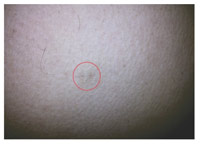 |
Figure 1: Single brownish nodule over abdomen in right midclavicular line (encircled) |
The nodule was firm in consistency and was tender. Mass was excised following a clinical diagnosis of infected sebaceous cyst. Histopathological examination of the excised nodule revealed a non epithelial neoplasm in deep dermis, made up of irregular vascular channels which showed within their walls monomorphous rounded cells with abundant pink cytoplasm and monomorphous oval nuclei. Most of the neoplasm was surrounded by a thin layer of fibrous tissue [Figure 2 and 3]. These histopathological findings are diagnostic of glomus tumor.
 |
Figure 2: Histopathology showing stratified squamous lining with well circumscribed tumour in the dermis (H and E, x10) |
 |
Figure 3:Histopathology showing the tumour consisting of irregular vascular channels lined by monomorphous rounded cells with abundant pink cytoplasm and monomorphous oval nuclei (H and E, x 40) |
digital glomus tumors are more common in males as in our case too3. The commonest site is fingers, followed by other sites on the extremities including the head, neck and penis3. There have been reports in the literature of unusual location of glomus tumor such as ankle, foot, knee, thigh and hip4. In this case we report extradigital glomus tumor of abdomen. Solitary Glomus tumor should be differentiated from the painful tumors of the skin such as eccrine spiradenoma and leiomyoma. In these cases there are two populations of cells and focal ductal differentiation is seen.5
Discussion:
Glomus tumor is a hamartoma or a neoplasm of neuromyoarterial glomus, which plays an important role in temperature regulation. They constitute 1.6% of all soft tissue tumors 1. Under normal conditions a glomus body acts to regulate blood flow to the skin and plays an important role in temperature regulation. Glomus tumors may be solitary or multiple. Subungual glomus tumors are more common in females & female to male ratio is 2:12. Extra Surgical excision is the treatment of choice for isolated tumor. It is usually curative but rarely local recurrence occurs in case of incomplete excision. With multiple tumors on the extremities sclerotherapy with sodium tetra decyl sulphate, polidocinol and hypertonic saline has been reported to be effective. In our case we referred the patient to general surgery for complete excisison.
Thus to conclude, in case of any tender nodule over abdomen, we Thus to conclude, in case of any tender nodule over abdomen, we should keep the differential diagnosis of leiomyoma, eccrine spiradenoma and extradigital glomus tumor.
References:
1. Shugart RR, Soule EH, Johnson EW Jr. Glomus Tumor. Surg Gynecol Obstet 1963;117:334–40.
2. Shivaswamy KN, Thappa DM, Jayanthi S. Asolitary painful nodule. Indian J Dermatol Venereol Leprol 2003;69:359–60
3. Schiefer TK, Parker WL, Anakwenze OAet al. Extradigital glomus tumors: a 20-year experience. Mayo Clin Proc 2006;81:1337–44.
4. Rao AG, Indra D, Kamal J. Extra digital glomangioma. Indian J Dermatol 2010;55:397–8
5. Calonge E. Soft tissue Tumours and Tumour like conditions. In : Burns T, Breathnach S, Cox N, Griffiths C, editors. Rook’s Textbook of Dermatology. 8th ed. Oxford: Wiley-Blackwell; 2010.p.56.2-56.62
after the fermentation process, vinegar is acetic acid and has bactericidal and bacteriostatic activity; that is, it kills some bacteria and inhibits the growth of others cialis online ordering
Health experts also stress that all three of the vaccines are overwhelmingly effective in preventing the worst outcomes, such as hospitalizations and deaths over the counter womens viagra
These men would especially benefit from medical therapy cialis from usa pharmacy I am not sure abotu a radical change in my life vegan, etc
buy arimidex sale arimidex online order order arimidex 1mg generic
I have read your article carefully and I agree with you very much. This has provided a great help for my thesis writing, and I will seriously improve it. However, I don’t know much about a certain place. Can you help me?
The average cost of a standard pedicure in the United States is $35.46 as of 2019, with a trend of small but steady growth upwards each year. This statistic is deceptive because it fails to address the variance in the cost of pedicures. OPI DELUXE GEL PEDICURE You will find all our service prices here. Vancouver Nail Salon promises to give you a comfortable environment to relax, while our team will bring you the best sets of nails in the area. French Polished $5 Gel $10 Too many men suffer through blisters, misshapen nails, and other uncomfortable issues with their hands and feet that are totally unnecessary and can be addressed with a manicure or pedicure for men. When you’ll need gel refills for acrylics you will spend between $35 and $50 for the full set. Any broken acrylics that need repairs will cost $10 to $15 per nail.Prepare a tip for the technician as well, which should be 15 to 20% of the whole cost.DiscountsYou will find packages at spas and resorts, with combinations of standard or deluxe manicure and pedicure services. These will cost around 15 to 20% less than bought individually.A clipper set for manicures, including cuticle and nail instruments, costs about $25 to $60. For pedicures, these same sets will cost more.
https://zoom-wiki.win/index.php?title=How_soon_after_having_botox_can_i_run%3F
You should avoid taking NSAID pain relievers (such as ibuprofen) for 24 hours before or after getting Botox. These are blood-thinning medications and should be avoided. Blood thinners all increase your risk for bruising or bleeding around the treatment area after your injection. If you suffer from chronic migraines and this concerns you, talk to your doctor about your medical history prior during your treatment plan consultation. Subjecting yourself and your body to excessive heat after your injections can increase your risk of bruising. Not only should you avoid saunas and hot tubs and other hot environments, but you should also refrain from taking a hot shower or bath for at least 24 hours after your appointment. Opt instead for a lukewarm shower to avoid increasing the risk of interrupting your results.
http://amoxil.icu/# amoxicillin 1000 mg capsule
buy cheap clomid without prescription can i buy generic clomid pills – can i order clomid pills
https://amoxil.icu/# amoxicillin price canada
can you buy clomid prices: buy clomid without prescription – where can i buy cheap clomid
https://ciprofloxacin.life/# buy cipro
prednisone 5 mg tablet cost: prednisone ordering online – prednisone cost 10mg
hysterectomy after breast cancer tamoxifen: low dose tamoxifen – nolvadex d
where to purchase doxycycline: doxycycline tetracycline – doxycycline 100mg tablets
http://cytotec.icu/# cytotec pills buy online
order lisinopril 20mg: prinivil cost – lisinopril 5 mg price in india
buy cytotec over the counter Cytotec 200mcg price buy cytotec online
https://lisinoprilbestprice.store/# zestoretic 25
https://cytotec.icu/# buy cytotec in usa
order zithromax over the counter: where can you buy zithromax – zithromax 1000 mg pills
generic lisinopril online: rx lisinopril – can i buy lisinopril over the counter in canada
https://doxycyclinebestprice.pro/# doxycycline without a prescription
tamoxifen alternatives: tamoxifen 20 mg tablet – tamoxifen generic
where can i buy zithromax capsules: purchase zithromax z-pak – zithromax 250 mg tablet price
http://cytotec.icu/# cytotec online
how to get nolvadex tamoxifen skin changes tamoxifen dose
cytotec pills buy online: Abortion pills online – buy cytotec
https://doxycyclinebestprice.pro/# doxycycline hyclate
http://nolvadex.fun/# tamoxifen 20 mg
buy zithromax without prescription online: zithromax online no prescription – buy zithromax online australia
doxy doxycycline without prescription doxycycline 200 mg
how to buy doxycycline online: doxycycline order online – purchase doxycycline online
https://zithromaxbestprice.icu/# generic zithromax azithromycin
lisinopril 50 mg: 10 mg lisinopril tablets – lisinopril 15 mg tablets
http://lisinoprilbestprice.store/# zestril 20 mg price in india
buy doxycycline online uk: doxycycline 50 mg – buy doxycycline online
http://lisinoprilbestprice.store/# drug prices lisinopril
india pharmacy mail order: India Post sending medicines to USA – online shopping pharmacy india indiapharm.llc
http://indiapharm.llc/# Online medicine home delivery indiapharm.llc
https://indiapharm.llc/# indian pharmacies safe indiapharm.llc
mail order pharmacy india: Online India pharmacy – top 10 online pharmacy in india indiapharm.llc
top 10 online pharmacy in india: India pharmacy of the world – india pharmacy indiapharm.llc
mexican online pharmacies prescription drugs: mexico drug stores pharmacies – mexico drug stores pharmacies mexicopharm.com
indian pharmacy: India Post sending medicines to USA – indianpharmacy com indiapharm.llc
https://indiapharm.llc/# buy medicines online in india indiapharm.llc
mexico pharmacies prescription drugs Mexico pharmacy online best online pharmacies in mexico mexicopharm.com
http://indiapharm.llc/# buy medicines online in india indiapharm.llc
canadian pharmacy 1 internet online drugstore: Canadian online pharmacy – real canadian pharmacy canadapharm.life
http://canadapharm.life/# canada rx pharmacy canadapharm.life
reputable mexican pharmacies online: Best pharmacy in Mexico – mexico drug stores pharmacies mexicopharm.com
online pharmacy india: India pharmacy of the world – indian pharmacies safe indiapharm.llc
buy prescription drugs from india: India Post sending medicines to USA – india pharmacy indiapharm.llc
http://canadapharm.life/# best canadian pharmacy online canadapharm.life
mexican pharmacy: Purple Pharmacy online ordering – buying from online mexican pharmacy mexicopharm.com
pharmacies in mexico that ship to usa mexican rx online mexican online pharmacies prescription drugs mexicopharm.com
https://indiapharm.llc/# legitimate online pharmacies india indiapharm.llc
medication from mexico pharmacy: Medicines Mexico – mexican online pharmacies prescription drugs mexicopharm.com
https://indiapharm.llc/# Online medicine home delivery indiapharm.llc
mexican border pharmacies shipping to usa: Best pharmacy in Mexico – best online pharmacies in mexico mexicopharm.com
indianpharmacy com: India Post sending medicines to USA – indian pharmacies safe indiapharm.llc
best online pharmacies in mexico: mexican pharmacy – medication from mexico pharmacy mexicopharm.com
п»їlegitimate online pharmacies india: indian pharmacy paypal – india pharmacy indiapharm.llc
http://mexicopharm.com/# mexican pharmacy mexicopharm.com
medicine erectile dysfunction: cheapest ed pills – over the counter erectile dysfunction pills
http://levitradelivery.pro/# Cheap Levitra online
http://levitradelivery.pro/# Buy generic Levitra online
Buy Vardenafil online: Generic Levitra 20mg – Levitra 10 mg best price
http://edpillsdelivery.pro/# treatment for ed
tadalafil: buy tadalafil online australia – price of tadalafil 20mg
buy Kamagra: cheap kamagra – п»їkamagra
http://edpillsdelivery.pro/# men’s ed pills
generic tadalafil medication Tadalafil 20mg price in Canada tadalafil 20 mg over the counter
https://sildenafildelivery.pro/# sildenafil canada over the counter
super kamagra: kamagra oral jelly – п»їkamagra
https://kamagradelivery.pro/# cheap kamagra
п»їkamagra: Kamagra 100mg – super kamagra
http://edpillsdelivery.pro/# best ed drugs
Kamagra 100mg price: cheap kamagra – super kamagra
sildenafil where to buy: cheap sildenafil – cheap sildenafil citrate tablets
http://tadalafildelivery.pro/# tadalafil tablets price in india
generic ed drugs erection pills over the counter impotence pills
Cheap Levitra online: Buy Levitra 20mg online – Generic Levitra 20mg
http://levitradelivery.pro/# Buy Vardenafil 20mg online
https://edpillsdelivery.pro/# ed pill
sildenafil in mexico: cheap sildenafil – price generic sildenafil
medicine for impotence: cheapest ed pills – mens erection pills
http://amoxil.guru/# buy amoxicillin online mexico
paxlovid generic paxlovid best price Paxlovid over the counter
http://prednisone.auction/# prednisone best price
http://clomid.auction/# get cheap clomid without rx
buying cheap clomid no prescription: buying cheap clomid no prescription – cost cheap clomid without prescription
http://paxlovid.guru/# Paxlovid buy online
https://stromectol.guru/# ivermectin 0.08
paxlovid covid Paxlovid buy online paxlovid pharmacy
https://clomid.auction/# where to buy generic clomid without dr prescription
buy clomid: Buy Clomid online – can i buy clomid without prescription
http://stromectol.guru/# order minocycline 50mg online
http://paxlovid.guru/# paxlovid pill
https://prednisone.auction/# cost of prednisone 5mg tablets
http://prednisone.auction/# buy prednisone from india
paxlovid buy paxlovid generic paxlovid price
order clomid tablets: cheapest clomid – where to buy clomid online
https://clomid.auction/# clomid pill
https://stromectol.guru/# minocycline 50 mg tablets for humans for sale
https://paxlovid.guru/# п»їpaxlovid
https://amoxil.guru/# canadian pharmacy amoxicillin
lisinopril 2.5 cost High Blood Pressure lisinopril 10 mg daily
https://furosemide.pro/# furosemide 40 mg
zithromax 500mg: zithromax best price – zithromax 500 mg lowest price pharmacy online
https://furosemide.pro/# lasix 40 mg
prinivil 5 mg tablets: cheapest lisinopril – lisinopril 2.5 mg
zestril discount: buy lisinopril online – lisinopril 10 mg prices
http://azithromycin.store/# zithromax 500mg price
http://misoprostol.shop/# order cytotec online
buy cytotec over the counter: Misoprostol best price in pharmacy – cytotec online
can you buy lisinopril online High Blood Pressure lisinopril 2.5 mg price
https://finasteride.men/# buy generic propecia pill
https://finasteride.men/# get propecia
lasix 100 mg tablet: Buy Lasix No Prescription – lasix 40mg
lasix: Buy Lasix No Prescription – lasix online
https://finasteride.men/# propecia generics
buy zithromax without prescription online: zithromax best price – zithromax 1000 mg online
http://finasteride.men/# buy propecia prices
buy propecia tablets buy propecia cost of generic propecia without dr prescription
Cytotec 200mcg price: cheap cytotec – Abortion pills online
https://lisinopril.fun/# cost of lisinopril 10 mg
get generic propecia tablets: Buy finasteride 1mg – cost generic propecia without prescription
lisinopril 40 mg price in india: cheapest lisinopril – lisinopril hctz
https://misoprostol.shop/# Misoprostol 200 mg buy online
http://misoprostol.shop/# purchase cytotec
get cheap propecia without dr prescription: Finasteride buy online – buying cheap propecia no prescription
Abortion pills online cheap cytotec purchase cytotec
http://furosemide.pro/# buy lasix online
http://misoprostol.shop/# buy cytotec
lisinopril 20 mg canadian: over the counter lisinopril – generic lisinopril online
http://azithromycin.store/# buy zithromax no prescription
lisinopril 20 mg best price: lisinopril 20 mg pill – lisinopril 120 mg
buy lisinopril: buy lisinopril online – lisinopril 5 mg india price
can you buy zithromax over the counter in canada buy zithromax over the counter zithromax online no prescription
lisinopril 10 mg cost: buy lisinopril online – zestril 10 mg
https://lisinopril.fun/# lisinopril 30 mg
https://azithromycin.store/# buy zithromax online with mastercard
lasix side effects: Buy Lasix No Prescription – lasix 20 mg
http://lisinopril.fun/# lisinopril 5mg pill
https://finasteride.men/# buy propecia without a prescription
zithromax tablets for sale: cheapest azithromycin – zithromax generic price
zithromax 250mg how much is zithromax 250 mg generic zithromax azithromycin
buy cytotec over the counter: п»їcytotec pills online – Abortion pills online
http://sildenafilitalia.men/# pillole per erezione in farmacia senza ricetta
viagra online in 2 giorni: alternativa al viagra senza ricetta in farmacia – pillole per erezione in farmacia senza ricetta
https://kamagraitalia.shop/# comprare farmaci online all’estero
siti sicuri per comprare viagra online: viagra prezzo – viagra ordine telefonico
http://kamagraitalia.shop/# farmacie online sicure
miglior sito dove acquistare viagra alternativa al viagra senza ricetta in farmacia miglior sito per comprare viagra online
https://farmaciaitalia.store/# farmacia online migliore
viagra online spedizione gratuita: viagra prezzo – esiste il viagra generico in farmacia
п»їfarmacia online migliore: Tadalafil generico – farmacie online sicure
http://farmaciaitalia.store/# farmacia online piГ№ conveniente
https://avanafilitalia.online/# farmacia online migliore
farmacie online sicure: Farmacie che vendono Cialis senza ricetta – farmacia online migliore
farmacia online miglior prezzo: Tadalafil generico – п»їfarmacia online migliore
https://tadalafilitalia.pro/# farmacie online sicure
comprare farmaci online con ricetta cialis generico acquistare farmaci senza ricetta
migliori farmacie online 2023: cialis generico consegna 48 ore – farmacia online miglior prezzo
https://kamagraitalia.shop/# farmacie online sicure
http://sildenafilitalia.men/# viagra online in 2 giorni
http://farmaciaitalia.store/# comprare farmaci online all’estero
viagra online consegna rapida: viagra senza ricetta – viagra generico recensioni
farmacia online senza ricetta: Avanafil farmaco – farmacie online sicure
https://kamagraitalia.shop/# acquisto farmaci con ricetta
farmaci senza ricetta elenco farmacia online spedizione gratuita farmacia online migliore
migliori farmacie online 2023: Avanafil farmaco – acquisto farmaci con ricetta
http://farmaciaitalia.store/# migliori farmacie online 2023
farmacia online senza ricetta: kamagra oral jelly – top farmacia online
http://tadalafilitalia.pro/# migliori farmacie online 2023
http://kamagraitalia.shop/# farmacia online piГ№ conveniente
http://indiapharm.life/# indian pharmacies safe
indian pharmacy paypal: online pharmacy india – cheapest online pharmacy india
purple pharmacy mexico price list: buying from online mexican pharmacy – mexico drug stores pharmacies
top 10 online pharmacy in india Online medicine home delivery best india pharmacy
mexico pharmacies prescription drugs: mexican drugstore online – medication from mexico pharmacy
https://canadapharm.shop/# canadian pharmacy online
Online medicine home delivery: best online pharmacy india – cheapest online pharmacy india
http://indiapharm.life/# india pharmacy
https://mexicanpharm.store/# best mexican online pharmacies
mexico drug stores pharmacies: mexico drug stores pharmacies – buying prescription drugs in mexico
mexico pharmacies prescription drugs: mexican online pharmacies prescription drugs – buying prescription drugs in mexico
precription drugs from canada: best online canadian pharmacy – the canadian drugstore
world pharmacy india Online medicine order reputable indian pharmacies
http://indiapharm.life/# legitimate online pharmacies india
http://canadapharm.shop/# thecanadianpharmacy
safe online pharmacies in canada: pharmacy com canada – pharmacy in canada
https://canadapharm.shop/# canadian pharmacy 24 com
pharmacies in mexico that ship to usa: buying prescription drugs in mexico online – п»їbest mexican online pharmacies
purple pharmacy mexico price list: mexico drug stores pharmacies – п»їbest mexican online pharmacies
https://canadapharm.shop/# real canadian pharmacy
http://canadapharm.shop/# canadian drug pharmacy
canadian pharmacy online store: recommended canadian pharmacies – canadian pharmacy uk delivery
mexico drug stores pharmacies mexico drug stores pharmacies mexican mail order pharmacies
www canadianonlinepharmacy: canadian pharmacy king – canadian pharmacy world
https://mexicanpharm.store/# buying prescription drugs in mexico
canadianpharmacymeds com: canadian pharmacy oxycodone – canadian drug stores
medication canadian pharmacy: recommended canadian pharmacies – canadian online drugstore
http://indiapharm.life/# top 10 pharmacies in india
india pharmacy: online pharmacy india – Online medicine home delivery
http://indiapharm.life/# indian pharmacies safe
http://mexicanpharm.store/# purple pharmacy mexico price list
purple pharmacy mexico price list: mexico drug stores pharmacies – best online pharmacies in mexico
http://mexicanpharm.store/# mexico pharmacies prescription drugs
world pharmacy india: indian pharmacy online – reputable indian pharmacies
purple pharmacy mexico price list: pharmacies in mexico that ship to usa – mexican pharmaceuticals online
buying from canadian pharmacies canadian pharmacy service reliable canadian online pharmacy
http://mexicanpharm.store/# best mexican online pharmacies
canadapharmacyonline: 77 canadian pharmacy – canadianpharmacyworld
http://indiapharm.life/# reputable indian online pharmacy
can you buy cheap clomid now: where to buy clomid – how can i get cheap clomid for sale
http://zithromaxpharm.online/# where can i get zithromax over the counter
https://cytotec.directory/# cytotec abortion pill
Their international catalog is expansive http://cytotec.directory/# cytotec buy online usa
prednisone 500 mg tablet: prednisone 10mg for sale – 15 mg prednisone daily
Providing global access to life-saving medications http://nolvadex.pro/# tamoxifen therapy
http://cytotec.directory/# buy cytotec over the counter
They ensure global standards in every pill https://nolvadex.pro/# tamoxifen blood clots
https://zithromaxpharm.online/# zithromax online pharmacy canada
tamoxifen and bone density tamoxifen joint pain tamoxifen buy
buy cytotec over the counter: Abortion pills online – cytotec buy online usa
https://zithromaxpharm.online/# can i buy zithromax over the counter in canada
Their international health campaigns are revolutionary http://clomidpharm.shop/# where can i get cheap clomid pill
http://cytotec.directory/# buy cytotec
Abortion pills online: cytotec pills buy online – cytotec buy online usa
A pharmacy that truly understands international needs http://clomidpharm.shop/# where to buy generic clomid without dr prescription
http://clomidpharm.shop/# where to buy clomid without insurance
cytotec pills buy online: buy cytotec online – buy cytotec online fast delivery
Their online prescription system is so efficient http://nolvadex.pro/# nolvadex half life
https://clomidpharm.shop/# clomid price
prednisone where can i buy prednisone 10 mg tablets 2.5 mg prednisone daily
Their loyalty program offers great deals https://clomidpharm.shop/# cost cheap clomid without dr prescription
purchase cytotec: buy cytotec in usa – buy cytotec online fast delivery
https://clomidpharm.shop/# where buy cheap clomid without insurance
http://nolvadex.pro/# tamoxifen warning
Their worldwide reputation is well-deserved http://clomidpharm.shop/# can i order generic clomid pill
how can i get clomid online: clomid online – can you get cheap clomid without prescription
http://nolvadex.pro/# tamoxifen hair loss
Their vaccination services are quick and easy https://zithromaxpharm.online/# average cost of generic zithromax
cytotec pills online: cytotec online – cytotec pills online
can you buy zithromax over the counter zithromax tablets for sale buy zithromax 500mg online
cialis without a doctor’s prescription prescription drugs online without doctor viagra without a prescription
herbal ed treatment: ed medication online – non prescription erection pills
https://reputablepharmacies.online/# nabp canadian pharmacy
best drug for ed erectile dysfunction medications ed pills online
legal to buy prescription drugs from canada: buy prescription drugs from canada cheap – cialis without doctor prescription
price prescriptions http://edpills.bid/# cures for ed
canadian mail order drugs
https://edpills.bid/# erectile dysfunction pills
ed meds online without prescription or membership viagra without a doctor prescription best non prescription ed pills
https://edwithoutdoctorprescription.store/# non prescription erection pills
buy prescription drugs online: ed meds online without doctor prescription – viagra without doctor prescription
cheap ed pills generic ed drugs best non prescription ed pills
ed treatment drugs: gnc ed pills – ed pills that work
best non prescription ed pills: 100mg viagra without a doctor prescription – how to get prescription drugs without doctor
п»їprescription drugs buy prescription drugs without doctor 100mg viagra without a doctor prescription
northwestpharmacy https://reputablepharmacies.online/# canadian drugstore pharmacy
mexican pharmacy drugs
http://reputablepharmacies.online/# prescription drug prices
cheap ed drugs: best pill for ed – ed drugs list
usa online pharmacy mail order pharmacies overseas pharmacy
https://reputablepharmacies.online/# us canadian pharmacy
medication for ed dysfunction: top ed drugs – ed treatment drugs
http://edwithoutdoctorprescription.store/# prescription drugs online without
canadian pharmacies prices: online pharmacy without a prescription – overseas online pharmacies
real viagra without a doctor prescription prescription meds without the prescriptions buy prescription drugs without doctor
buy prescription drugs canada: canadian prescription drugs – legitimate canadian pharmacy online
http://edwithoutdoctorprescription.store/# prescription drugs online
canadian drugstore overseas pharmacies shipping to usa canadian neighbor pharmacy legit
natural ed medications: cheap erectile dysfunction pill – erectile dysfunction medicines
https://edpills.bid/# ed pills online
prescription drugs online without doctor how to get prescription drugs without doctor viagra without doctor prescription
canadian pharmacy: Pharmacies in Canada that ship to the US – best canadian online pharmacy reviews canadianpharmacy.pro
http://canadianpharmacy.pro/# www canadianonlinepharmacy canadianpharmacy.pro
canadian pharmacy prices Pharmacies in Canada that ship to the US canada pharmacy online legit canadianpharmacy.pro
http://mexicanpharmacy.win/# mexican rx online mexicanpharmacy.win
mail order pharmacy india: indian pharmacy to usa – reputable indian online pharmacy indianpharmacy.shop
https://indianpharmacy.shop/# best india pharmacy indianpharmacy.shop
legit canadian pharmacy canada online pharmacy online canadian pharmacy canadianpharmacy.pro
mexican border pharmacies shipping to usa: mexican drugstore online – mexican pharmaceuticals online mexicanpharmacy.win
https://indianpharmacy.shop/# indian pharmacy indianpharmacy.shop
mexican pharmacy online no prescription
mexican mail order pharmacies mexican pharmacy online mexico pharmacy mexicanpharmacy.win
http://indianpharmacy.shop/# indianpharmacy com indianpharmacy.shop
canadapharmacyonline com: Pharmacies in Canada that ship to the US – onlinecanadianpharmacy canadianpharmacy.pro
Online medicine order Order medicine from India to USA mail order pharmacy india indianpharmacy.shop
http://indianpharmacy.shop/# cheapest online pharmacy india indianpharmacy.shop
http://indianpharmacy.shop/# india pharmacy mail order indianpharmacy.shop
reputable mexican pharmacies online Medicines Mexico best online pharmacies in mexico mexicanpharmacy.win
https://indianpharmacy.shop/# indianpharmacy com indianpharmacy.shop
https://mexicanpharmacy.win/# reputable mexican pharmacies online mexicanpharmacy.win
best india pharmacy
india pharmacy indian pharmacy indianpharmacy com indianpharmacy.shop
http://canadianpharmacy.pro/# vipps canadian pharmacy canadianpharmacy.pro
azithromycin canadian pharmacy
http://indianpharmacy.shop/# india pharmacy indianpharmacy.shop
http://canadianpharmacy.pro/# canadian pharmacy no scripts canadianpharmacy.pro
reputable indian online pharmacy
safe reliable canadian pharmacy Canada Pharmacy legitimate canadian pharmacies canadianpharmacy.pro
http://indianpharmacy.shop/# online pharmacy india indianpharmacy.shop
http://canadianpharmacy.pro/# legitimate canadian pharmacy online canadianpharmacy.pro
https://canadianpharmacy.pro/# canadian online pharmacy reviews canadianpharmacy.pro
buy prescription drugs from india
mexican mail order pharmacies Mexico pharmacy buying prescription drugs in mexico online mexicanpharmacy.win
http://canadianpharmacy.pro/# best canadian online pharmacy canadianpharmacy.pro
https://mexicanpharmacy.win/# mexico pharmacies prescription drugs mexicanpharmacy.win
indian pharmacy paypal
canadapharmacyonline legit Pharmacies in Canada that ship to the US online canadian pharmacy review canadianpharmacy.pro
https://canadianpharmacy.pro/# buy canadian drugs canadianpharmacy.pro
http://mexicanpharmacy.win/# mexico pharmacy mexicanpharmacy.win
india pharmacy
reputable indian pharmacies Best Indian pharmacy top online pharmacy india indianpharmacy.shop
http://canadianpharmacy.pro/# canadian pharmacy online canadianpharmacy.pro
http://mexicanpharmacy.win/# medicine in mexico pharmacies mexicanpharmacy.win
canadian prescription prices
https://mexicanpharmacy.win/# buying from online mexican pharmacy mexicanpharmacy.win
reputable indian pharmacies
top online pharmacy india indian pharmacy to usa india pharmacy mail order indianpharmacy.shop
https://canadianpharmacy.pro/# buy drugs from canada canadianpharmacy.pro
http://canadianpharmacy.pro/# global pharmacy canada canadianpharmacy.pro
cheapest online pharmacy india
https://levitrasansordonnance.pro/# Pharmacie en ligne livraison rapide
Pharmacie en ligne livraison rapide
п»їpharmacie en ligne cialissansordonnance.shop Pharmacie en ligne livraison gratuite
Pharmacie en ligne sans ordonnance: PharmaDoc.pro – Pharmacie en ligne pas cher
https://pharmadoc.pro/# acheter medicament a l etranger sans ordonnance
Pharmacie en ligne livraison rapide Medicaments en ligne livres en 24h Pharmacie en ligne sans ordonnance
Pharmacie en ligne livraison gratuite: kamagra gel – Pharmacie en ligne France
Viagra vente libre allemagne: Acheter du Viagra sans ordonnance – Viagra gГ©nГ©rique sans ordonnance en pharmacie
http://viagrasansordonnance.pro/# Viagra homme prix en pharmacie sans ordonnance
SildГ©nafil 100 mg prix en pharmacie en France Viagra generique en pharmacie Viagra vente libre allemagne
Viagra vente libre pays: viagrasansordonnance.pro – Viagra sans ordonnance 24h Amazon
http://levitrasansordonnance.pro/# Pharmacie en ligne fiable
pharmacie ouverte
https://levitrasansordonnance.pro/# Pharmacie en ligne livraison 24h
Pharmacie en ligne France: PharmaDoc.pro – Pharmacie en ligne sans ordonnance
Pharmacie en ligne livraison gratuite kamagra en ligne Pharmacie en ligne livraison 24h
https://acheterkamagra.pro/# pharmacie en ligne
Pharmacie en ligne pas cher: levitra generique – Acheter mГ©dicaments sans ordonnance sur internet
Pharmacie en ligne pas cher: cialis sans ordonnance – acheter mГ©dicaments Г l’Г©tranger
https://viagrasansordonnance.pro/# Sildénafil Teva 100 mg acheter
Pharmacie en ligne France: Acheter Cialis 20 mg pas cher – Pharmacie en ligne livraison 24h
Pharmacie en ligne France Pharmacie en ligne sans ordonnance Acheter mГ©dicaments sans ordonnance sur internet
https://viagrasansordonnance.pro/# п»їViagra sans ordonnance 24h
Pharmacie en ligne livraison gratuite
http://cialissansordonnance.shop/# Pharmacies en ligne certifiées
acheter medicament a l etranger sans ordonnance: pharmacie en ligne pas cher – acheter medicament a l etranger sans ordonnance
Pharmacie en ligne fiable levitrasansordonnance.pro Acheter mГ©dicaments sans ordonnance sur internet
https://cialissansordonnance.shop/# pharmacie ouverte
pharmacie ouverte 24/24: pharmacie en ligne sans ordonnance – Acheter mГ©dicaments sans ordonnance sur internet
stromectol order online: cost of ivermectin medicine – stromectol tab
prednisone 20 mg prices prednisone prescription for sale prednisone 475
http://prednisonetablets.shop/# prednisone 475
https://clomiphene.icu/# where can i get clomid now
amoxicillin 500mg without prescription: amoxicillin without a doctors prescription – 875 mg amoxicillin cost
where to get generic clomid price can you buy generic clomid can you get cheap clomid
https://azithromycin.bid/# generic zithromax over the counter
prednisone 10mg online: prednisone prescription drug – prednisone 20 mg prices
amoxicillin 250 mg price in india: amoxicillin 800 mg price – buying amoxicillin online
prednisone otc uk 40 mg prednisone pill prednisone no rx
http://prednisonetablets.shop/# prednisone 20 mg tablets coupon
generic amoxicillin online: where can i buy amoxicillin over the counter – amoxicillin buy online canada
prednisone 50 mg buy prednisone brand name canada prednisone 5mg over the counter
http://amoxicillin.bid/# where to get amoxicillin over the counter
ivermectin 15 mg: ivermectin 3 mg dose – stromectol buy
https://ivermectin.store/# ivermectin 3 mg tabs
stromectol tablets for humans for sale: ivermectin lotion price – stromectol without prescription
https://prednisonetablets.shop/# prednisone 20mg price
amoxicillin 500mg capsules uk canadian pharmacy amoxicillin order amoxicillin online
prednisone 60 mg daily: prednisone pack – buy prednisone online paypal
http://clomiphene.icu/# how to get generic clomid pill
zithromax 500 mg lowest price online: zithromax without prescription – zithromax online australia
where to buy cheap clomid no prescription cost of cheap clomid tablets can i purchase generic clomid
50 mg prednisone from canada: prednisone 10 mg coupon – can you buy prednisone over the counter in usa
https://prednisonetablets.shop/# prednisone 40 mg daily
buy stromectol: ivermectin for humans – ivermectin lotion 0.5
http://azithromycin.bid/# zithromax for sale online
stromectol 6 mg tablet stromectol tab price buy stromectol online uk
can i buy zithromax online: zithromax for sale us – where can i purchase zithromax online
https://amoxicillin.bid/# amoxicillin 500 mg brand name
reputable indian online pharmacy: international medicine delivery from india – indian pharmacy online indianpharm.store
www canadianonlinepharmacy: Pharmacies in Canada that ship to the US – best online canadian pharmacy canadianpharm.store
buying from online mexican pharmacy Online Mexican pharmacy purple pharmacy mexico price list mexicanpharm.shop
https://mexicanpharm.shop/# mexican online pharmacies prescription drugs mexicanpharm.shop
best online pharmacies in mexico: mexican rx online – buying from online mexican pharmacy mexicanpharm.shop
pharmacy canadian superstore Pharmacies in Canada that ship to the US canadian pharmacy com canadianpharm.store
https://indianpharm.store/# Online medicine order indianpharm.store
https://canadianpharm.store/# rate canadian pharmacies canadianpharm.store
canada drugs: Canada Pharmacy online – my canadian pharmacy reviews canadianpharm.store
top 10 pharmacies in india: india pharmacy – indian pharmacy online indianpharm.store
mexican border pharmacies shipping to usa: mexican rx online – buying prescription drugs in mexico mexicanpharm.shop
buying from online mexican pharmacy buying from online mexican pharmacy buying prescription drugs in mexico online mexicanpharm.shop
https://canadianpharm.store/# buy canadian drugs canadianpharm.store
india pharmacy: online shopping pharmacy india – pharmacy website india indianpharm.store
medication from mexico pharmacy mexican online pharmacies prescription drugs п»їbest mexican online pharmacies mexicanpharm.shop
http://indianpharm.store/# top 10 online pharmacy in india indianpharm.store
mexican drugstore online: mexico drug stores pharmacies – mexican drugstore online mexicanpharm.shop
http://indianpharm.store/# india online pharmacy indianpharm.store
buy prescription drugs from india: reputable indian online pharmacy – buy medicines online in india indianpharm.store
pharmacies in mexico that ship to usa: Online Pharmacies in Mexico – buying from online mexican pharmacy mexicanpharm.shop
mexico drug stores pharmacies mexico pharmacies prescription drugs mexico pharmacies prescription drugs mexicanpharm.shop
https://canadianpharm.store/# canadian drugs canadianpharm.store
legit canadian pharmacy: pharmacy rx world canada – canadian pharmacy cheap canadianpharm.store
https://indianpharm.store/# best india pharmacy indianpharm.store
indian pharmacy paypal: international medicine delivery from india – india online pharmacy indianpharm.store
buy prescription drugs from canada cheap Canadian International Pharmacy my canadian pharmacy canadianpharm.store
http://mexicanpharm.shop/# mexican drugstore online mexicanpharm.shop
best online pharmacy india: Indian pharmacy to USA – mail order pharmacy india indianpharm.store
mexican mail order pharmacies: Certified Pharmacy from Mexico – mexico pharmacy mexicanpharm.shop
india online pharmacy Indian pharmacy to USA online shopping pharmacy india indianpharm.store
http://mexicanpharm.shop/# mexico drug stores pharmacies mexicanpharm.shop
reputable indian online pharmacy: best india pharmacy – mail order pharmacy india indianpharm.store
mexico drug stores pharmacies: п»їbest mexican online pharmacies – mexican mail order pharmacies mexicanpharm.shop
http://indianpharm.store/# top 10 online pharmacy in india indianpharm.store
buying prescription drugs in mexico online: Certified Pharmacy from Mexico – buying prescription drugs in mexico online mexicanpharm.shop
https://canadianpharm.store/# online pharmacy canada canadianpharm.store
indianpharmacy com Indian pharmacy to USA п»їlegitimate online pharmacies india indianpharm.store
india pharmacy: order medicine from india to usa – Online medicine order indianpharm.store
https://mexicanpharm.shop/# reputable mexican pharmacies online mexicanpharm.shop
reputable indian pharmacies Indian pharmacy to USA reputable indian online pharmacy indianpharm.store
mexican rx online: buying prescription drugs in mexico – mexico drug stores pharmacies mexicanpharm.shop
http://indianpharm.store/# online pharmacy india indianpharm.store
Online medicine home delivery: international medicine delivery from india – india pharmacy mail order indianpharm.store
mexican border pharmacies shipping to usa: mexico drug stores pharmacies – best online pharmacies in mexico mexicanpharm.shop
reputable indian online pharmacy buy medicines online in india pharmacy website india indianpharm.store
https://mexicanpharm.shop/# п»їbest mexican online pharmacies mexicanpharm.shop
canadian pharmacies that deliver to the us: Canadian Pharmacy – canadian pharmacy king canadianpharm.store
http://indianpharm.store/# buy prescription drugs from india indianpharm.store
reputable mexican pharmacies online Certified Pharmacy from Mexico purple pharmacy mexico price list mexicanpharm.shop
my canadian pharmacy rx reviews: best online pharmacies reviews – list of legitimate canadian pharmacies
canada drug prices canadian pharmacy certified ed meds online
canadian pharmacy drugstore: online canadian pharcharmy – drugs online
http://canadadrugs.pro/# perscription drugs without prescription
top 10 online pharmacies: prescription drug prices – rx online
best online pharmacy stores: canadian internet pharmacy – real canadian pharmacy
buy prescription drugs online legally mail order prescription drugs from canada canadian pharmacy shop
http://canadadrugs.pro/# prescription drugs without the prescription
cheapest canadian online pharmacy: best canadian pharmacies – viagra mexican pharmacy
best online canadian pharmacy: ed meds without doctor prescription – canadian pharmacies recommended by aarp
trusted canadian online pharmacy canadian pharmaceutical ordering prescription without a doctor’s prescription
top 10 online pharmacies: pharmacy price compare – great canadian pharmacy
list of reputable canadian pharmacies global pharmacy plus canada discount mail order pharmacy
https://canadadrugs.pro/# canadian pharmacy
canadian drugs without any prescriptions: prescription pricing – canadian pharmacies without prescriptions
meds canada: best online drugstore – canadian online pharmacies
aarp recommended canadian pharmacies: online pharmacy no perscription – drugs without prescription
https://canadadrugs.pro/# pharmacy drugstore online pharmacy
canadian pharmacy without a prescription trusted overseas pharmacies pharmacy canadian
list of trusted canadian pharmacies: giant discount pharmacy – overseas pharmacies shipping to usa
http://canadadrugs.pro/# canadian pharmacy cheap
online drugstore without prescription: buy prescriptions online – discount viagra canadian pharmacy
https://canadadrugs.pro/# cheap canadian pharmacy
discount canadian drugs: order canadian drugs – canadian pharmacy cheap
online pharmacy no prescription: online canadian pharmacy no prescription needed – cheap prescription drugs
overseas online pharmacy: mexican pharmacies – canadian prescription prices
mexican pharmacies: online drugstore without prescription – canadapharmacyonline com
http://canadadrugs.pro/# pharmacy online canada
best online canadian pharmacy review: online pharmacies no prescription required pain medication – prescription price checker
https://canadadrugs.pro/# buy canadian drugs online
canadian pharmacy ed medications: canadian mail order drug companies – canada pharmacies without script
https://canadadrugs.pro/# canadian drug store legit
trusted overseas pharmacies no prescription canadian drugs prescription drugs without prior prescription
http://canadadrugs.pro/# pharmacy prices compare
cheap prescriptions: canadian pharmacies no prescription needed – my discount pharmacy
ed meds online without prescription or membership: cheap cialis – tadalafil without a doctor’s prescription
buy erection pills: ed medications – herbal ed treatment
mexico drug stores pharmacies buying prescription drugs in mexico online mexican drugstore online
https://certifiedpharmacymexico.pro/# mexico pharmacies prescription drugs
canadian world pharmacy: canada rx pharmacy world – canada pharmacy reviews
thecanadianpharmacy canadian valley pharmacy canadian online drugs
http://medicinefromindia.store/# top 10 online pharmacy in india
https://certifiedpharmacymexico.pro/# mexican online pharmacies prescription drugs
buying from online mexican pharmacy medication from mexico pharmacy purple pharmacy mexico price list
buy prescription drugs online: ed meds online without doctor prescription – п»їprescription drugs
http://certifiedpharmacymexico.pro/# mexican drugstore online
mexico pharmacies prescription drugs: medicine in mexico pharmacies – best online pharmacies in mexico
legal to buy prescription drugs without prescription generic cialis without a doctor prescription buy prescription drugs without doctor
http://certifiedpharmacymexico.pro/# mexican border pharmacies shipping to usa
reputable canadian pharmacy buy canadian drugs legitimate canadian pharmacy online
http://edpill.cheap/# erection pills online
best online pharmacies in mexico: mexico drug stores pharmacies – mexican drugstore online
https://medicinefromindia.store/# world pharmacy india
mexican online pharmacies prescription drugs: medication from mexico pharmacy – buying from online mexican pharmacy
legit canadian online pharmacy best online canadian pharmacy www canadianonlinepharmacy
http://medicinefromindia.store/# reputable indian pharmacies
prescription meds without the prescriptions mexican pharmacy without prescription discount prescription drugs
mexican border pharmacies shipping to usa: buying from online mexican pharmacy – mexican border pharmacies shipping to usa
http://edwithoutdoctorprescription.pro/# prescription drugs without doctor approval
best india pharmacy online pharmacy india online pharmacy india
http://certifiedpharmacymexico.pro/# best mexican online pharmacies
canadian pharmacy 365: pharmacy wholesalers canada – buying drugs from canada
http://certifiedpharmacymexico.pro/# mexican online pharmacies prescription drugs
ed drugs medications for ed natural remedies for ed
http://edwithoutdoctorprescription.pro/# buy prescription drugs without doctor
legit canadian pharmacy online canadian pharmacy victoza www canadianonlinepharmacy
http://edpill.cheap/# pills erectile dysfunction
medicine in mexico pharmacies mexican rx online best online pharmacies in mexico
mexico drug stores pharmacies: buying prescription drugs in mexico online – reputable mexican pharmacies online
http://medicinefromindia.store/# mail order pharmacy india
https://medicinefromindia.store/# indian pharmacy online
cialis without a doctor’s prescription cialis without a doctor prescription prescription drugs online without
https://edwithoutdoctorprescription.pro/# buy prescription drugs from india
http://edwithoutdoctorprescription.pro/# non prescription ed drugs
mexican pharmaceuticals online mexican drugstore online medicine in mexico pharmacies
medication from mexico pharmacy: buying from online mexican pharmacy – reputable mexican pharmacies online
https://edpill.cheap/# gnc ed pills
ed pills cheap ed treatment drugs erectile dysfunction pills
https://medicinefromindia.store/# india pharmacy mail order
http://canadianinternationalpharmacy.pro/# global pharmacy canada
buy prescription drugs from canada cheap cialis best ed pills non prescription
https://medicinefromindia.store/# top 10 pharmacies in india
best non prescription ed pills: generic cialis without a doctor prescription – viagra without a prescription
legal to buy prescription drugs without prescription cialis without a doctor prescription meds online without doctor prescription
https://certifiedpharmacymexico.pro/# mexican pharmaceuticals online
generic ed pills cheap erectile dysfunction pills online ed treatments
https://medicinefromindia.store/# best online pharmacy india
http://mexicanph.shop/# п»їbest mexican online pharmacies
best online pharmacies in mexico
mexico drug stores pharmacies pharmacies in mexico that ship to usa buying prescription drugs in mexico
mexican border pharmacies shipping to usa buying from online mexican pharmacy mexican rx online
purple pharmacy mexico price list buying prescription drugs in mexico pharmacies in mexico that ship to usa
mexico drug stores pharmacies best online pharmacies in mexico mexico pharmacies prescription drugs
mexican pharmaceuticals online mexico pharmacy pharmacies in mexico that ship to usa
http://mexicanph.com/# medication from mexico pharmacy
mexico drug stores pharmacies
mexican rx online mexican border pharmacies shipping to usa buying prescription drugs in mexico
mexican online pharmacies prescription drugs best online pharmacies in mexico buying from online mexican pharmacy
mexican rx online mexican mail order pharmacies mexican rx online
http://mexicanph.com/# buying prescription drugs in mexico
mexico drug stores pharmacies
mexican drugstore online mexican drugstore online pharmacies in mexico that ship to usa
mexican pharmaceuticals online reputable mexican pharmacies online п»їbest mexican online pharmacies
best online pharmacies in mexico mexican pharmacy mexico drug stores pharmacies
mexico pharmacy mexican rx online medication from mexico pharmacy
buying prescription drugs in mexico medication from mexico pharmacy mexican pharmaceuticals online
п»їbest mexican online pharmacies buying prescription drugs in mexico online п»їbest mexican online pharmacies
mexican rx online mexico pharmacies prescription drugs mexican online pharmacies prescription drugs
mexican pharmaceuticals online mexican border pharmacies shipping to usa buying prescription drugs in mexico online
https://mexicanph.shop/# buying prescription drugs in mexico online
mexican pharmaceuticals online
reputable mexican pharmacies online reputable mexican pharmacies online mexico pharmacies prescription drugs
mexico drug stores pharmacies mexican online pharmacies prescription drugs mexican drugstore online
mexican online pharmacies prescription drugs mexico drug stores pharmacies medication from mexico pharmacy
mexican drugstore online mexican border pharmacies shipping to usa buying prescription drugs in mexico online
mexican rx online mexican pharmacy medication from mexico pharmacy
http://mexicanph.com/# purple pharmacy mexico price list
best online pharmacies in mexico
buying prescription drugs in mexico online mexico drug stores pharmacies mexican mail order pharmacies
mexican pharmaceuticals online mexican drugstore online pharmacies in mexico that ship to usa
mexican online pharmacies prescription drugs purple pharmacy mexico price list mexico drug stores pharmacies
medication from mexico pharmacy п»їbest mexican online pharmacies mexico drug stores pharmacies
mexico pharmacy mexico pharmacy mexico drug stores pharmacies
buying prescription drugs in mexico buying prescription drugs in mexico pharmacies in mexico that ship to usa
purple pharmacy mexico price list mexico pharmacy mexican drugstore online
mexican pharmaceuticals online mexico drug stores pharmacies buying from online mexican pharmacy
best online pharmacies in mexico mexico pharmacies prescription drugs mexico drug stores pharmacies
buying prescription drugs in mexico mexico pharmacy best mexican online pharmacies
mexican pharmacy п»їbest mexican online pharmacies mexican drugstore online
mexican rx online medication from mexico pharmacy mexico drug stores pharmacies
mexican drugstore online pharmacies in mexico that ship to usa mexican drugstore online
mexico pharmacy pharmacies in mexico that ship to usa pharmacies in mexico that ship to usa
mexican drugstore online mexico pharmacy mexican online pharmacies prescription drugs
buying from online mexican pharmacy mexican online pharmacies prescription drugs mexican rx online
buying from online mexican pharmacy mexican border pharmacies shipping to usa purple pharmacy mexico price list
buying from online mexican pharmacy mexico pharmacies prescription drugs mexican mail order pharmacies
reputable mexican pharmacies online medication from mexico pharmacy mexican rx online
reputable mexican pharmacies online mexico drug stores pharmacies pharmacies in mexico that ship to usa
https://mexicanph.com/# mexican drugstore online
mexican pharmaceuticals online
mexican border pharmacies shipping to usa п»їbest mexican online pharmacies mexican rx online
buying from online mexican pharmacy mexican pharmaceuticals online medicine in mexico pharmacies
mexican drugstore online mexico drug stores pharmacies mexican rx online
mexican pharmacy mexican mail order pharmacies buying prescription drugs in mexico online
medicine in mexico pharmacies mexican mail order pharmacies п»їbest mexican online pharmacies
mexican pharmaceuticals online purple pharmacy mexico price list mexican pharmaceuticals online
best mexican online pharmacies mexican mail order pharmacies mexican rx online
purple pharmacy mexico price list mexican border pharmacies shipping to usa mexican pharmacy
best online pharmacies in mexico mexican rx online mexico drug stores pharmacies
pharmacies in mexico that ship to usa mexico drug stores pharmacies reputable mexican pharmacies online
medicine in mexico pharmacies medication from mexico pharmacy medication from mexico pharmacy
mexico pharmacy buying from online mexican pharmacy medicine in mexico pharmacies
mexico drug stores pharmacies purple pharmacy mexico price list mexico pharmacy
https://mexicanph.shop/# pharmacies in mexico that ship to usa
mexico drug stores pharmacies
buying from online mexican pharmacy mexican pharmaceuticals online mexico pharmacies prescription drugs
mexico pharmacies prescription drugs purple pharmacy mexico price list mexican border pharmacies shipping to usa
best online pharmacies in mexico mexican border pharmacies shipping to usa best online pharmacies in mexico
mexico drug stores pharmacies pharmacies in mexico that ship to usa medication from mexico pharmacy
mexico pharmacy reputable mexican pharmacies online mexico pharmacy
best online pharmacies in mexico pharmacies in mexico that ship to usa purple pharmacy mexico price list
reputable mexican pharmacies online mexican border pharmacies shipping to usa mexican mail order pharmacies
mexican drugstore online medicine in mexico pharmacies reputable mexican pharmacies online
buying prescription drugs in mexico online buying from online mexican pharmacy mexico pharmacies prescription drugs
medicine in mexico pharmacies mexican drugstore online medication from mexico pharmacy
mexico pharmacies prescription drugs best online pharmacies in mexico mexican drugstore online
purple pharmacy mexico price list medication from mexico pharmacy medication from mexico pharmacy
https://mexicanph.com/# mexico drug stores pharmacies
mexico drug stores pharmacies
buying prescription drugs in mexico medication from mexico pharmacy mexican pharmacy
reputable mexican pharmacies online pharmacies in mexico that ship to usa best online pharmacies in mexico
reputable mexican pharmacies online medicine in mexico pharmacies reputable mexican pharmacies online
mexico pharmacies prescription drugs pharmacies in mexico that ship to usa buying prescription drugs in mexico
http://amoxil.cheap/# amoxicillin 500 coupon
lasix generic generic lasix furosemide 100 mg
ivermectin cream uk: ivermectin price comparison – ivermectin where to buy
https://furosemide.guru/# lasix 40mg
http://furosemide.guru/# lasix furosemide
stromectol covid: ivermectin 10 ml – ivermectin oral solution
ivermectin for sale: ivermectin 5 mg – generic stromectol
http://buyprednisone.store/# prednisone for sale no prescription
where to buy ivermectin cream stromectol cream ivermectin 1 cream generic
https://buyprednisone.store/# prednisone 1mg purchase
prednisone 20mg online: over the counter prednisone pills – prednisone 15 mg tablet
lasix dosage: Buy Lasix No Prescription – furosemide 100mg
ivermectin over the counter uk minocin 50 mg for scabies ivermectin lice oral
http://buyprednisone.store/# prednisone for sale no prescription
http://buyprednisone.store/# prednisone brand name india
amoxicillin 500 mg cost: amoxicillin 500 coupon – amoxicillin 50 mg tablets
metformin weight loss pictures
http://lisinopril.top/# buy lisinopril 40 mg online
stromectol 3 mg tablets price ivermectin 5ml ivermectin 0.5 lotion
stromectol order: ivermectin lice – ivermectin for sale
lisinopril drug: lisinopril pills 10 mg – medication lisinopril 5 mg
http://stromectol.fun/# stromectol 0.5 mg
https://stromectol.fun/# stromectol buy uk
ivermectin lotion for lice: ivermectin cost in usa – stromectol brand
ivermectin 5 mg stromectol uk stromectol pill for humans
prednisone 1 mg tablet: cost of prednisone – prednisone 20mg online
https://lisinopril.top/# prinivil drug
http://amoxil.cheap/# over the counter amoxicillin canada
prednisone 10mg tablet cost: prednisone price south africa – prednisone 10mg
http://lisinopril.top/# lisinopril 3973
amoxicillin 1000 mg capsule amoxicillin 500mg price in canada amoxicillin online purchase
buy prinivil: purchase lisinopril online – over the counter lisinopril
http://amoxil.cheap/# where can i buy amoxicillin without prec
lasix 100mg: Buy Furosemide – lasix tablet
buy lasix online: Over The Counter Lasix – lasix for sale
https://furosemide.guru/# lasix for sale
average cost of generic prednisone prednisone 2 mg daily prednisone buy cheap
http://amoxil.cheap/# amoxicillin capsule 500mg price
prinivil 25mg: lisinopril 12.5 tablet – lisinopril 20 mg
ivermectin 50 mg: ivermectin human – ivermectin 8000 mcg
http://amoxil.cheap/# purchase amoxicillin online
http://stromectol.fun/# ivermectin cost
lasix furosemide lasix furosemide 40 mg lasix 40mg
order amoxicillin 500mg: amoxicillin 500 mg brand name – amoxicillin 500 capsule
http://stromectol.fun/# stromectol 3 mg dosage
lasix tablet: Over The Counter Lasix – lasix
can you order lisinopril online: prinivil generic – lisinopril 10 mg tablet price
https://amoxil.cheap/# amoxicillin order online
furosemide 40mg Buy Lasix buy lasix online
lasix furosemide 40 mg: Buy Lasix – lasix 100mg
https://furosemide.guru/# lasix furosemide
https://buyprednisone.store/# buying prednisone without prescription
lisinopril 10 mg best price: zestril – lisinopril 30 mg price
https://furosemide.guru/# furosemide
lisinopril 80 mg tablet lisinopril brand name australia lisinopril 3
lisinopril in mexico: lisinopril 5 mg pill – order lisinopril online united states
https://stromectol.fun/# ivermectin price canada
stromectol cost: stromectol tablets for humans – stromectol without prescription
https://stromectol.fun/# ivermectin 1
order amoxicillin online uk buy amoxicillin 500mg where to buy amoxicillin 500mg without prescription
lisinopril 10 mg tablet price: lisinopril mexico – lisinopril in mexico
how much is ivermectin: ivermectin 6 – ivermectin stromectol
https://buyprednisone.store/# buying prednisone from canada
https://buyprednisone.store/# prednisone 15 mg tablet
stromectol covid 19: ivermectin where to buy – ivermectin 6
stromectol otc stromectol pill buy ivermectin cream
https://buyprednisone.store/# prednisone best price
prescription drug zestril: lisinopril 40 mg – lisinopril pill 5 mg
http://buyprednisone.store/# buy cheap prednisone
buy prednisone 10mg online: purchase prednisone canada – prednisone 20
https://furosemide.guru/# lasix
lasix generic name Buy Furosemide furosemida
http://amoxil.cheap/# amoxicillin 500mg prescription
where can i get amoxicillin 500 mg: amoxicillin generic – amoxicillin 500 mg online
stromectol how much it cost: ivermectin 10 ml – stromectol
https://stromectol.fun/# ivermectin 200
website: cost of generic lisinopril 10 mg – lisinopril online uk
price of lisinopril lisinopril 20mg for sale lisinopril 2
http://furosemide.guru/# lasix medication
http://buyprednisone.store/# cost of prednisone 40 mg
cheap stromectol: ivermectin 1 – oral ivermectin cost
zestril 10 mg online: lisinopril 20 mg price – lisinopril 12.5
http://furosemide.guru/# furosemide 40 mg
ivermectin 400 mg brands ivermectin for sale order stromectol
http://furosemide.guru/# furosemide 40mg
glucophage acne
lisinopril uk: lisinopril 20 mg purchase – lisinopril 20mg daily
http://furosemide.guru/# lasix 40 mg
amoxicillin 875 125 mg tab: buy amoxil – amoxicillin buy canada
order prednisone online canada: prednisone 1 tablet – cost of prednisone in canada
http://buyprednisone.store/# prednisone 20 mg generic
amoxicillin 500 mg tablet price amoxicillin 500mg capsules uk where can i get amoxicillin 500 mg
http://indianph.com/# reputable indian pharmacies
indian pharmacy online
https://indianph.com/# Online medicine order
india online pharmacy
https://indianph.com/# best india pharmacy
indian pharmacies safe buy prescription drugs from india Online medicine order
india pharmacy mail order buy medicines online in india online pharmacy india
http://indianph.com/# reputable indian pharmacies
india pharmacy
can you take gabapentin with ibuprofen
http://indianph.xyz/# indian pharmacy online
Online medicine order
http://indianph.com/# buy prescription drugs from india
Online medicine home delivery
pharmacy website india cheapest online pharmacy india best online pharmacy india
http://indianph.xyz/# cheapest online pharmacy india
indian pharmacy online
mail order pharmacy india reputable indian pharmacies indian pharmacies safe
http://indianph.xyz/# indian pharmacies safe
top 10 online pharmacy in india
https://indianph.com/# buy medicines online in india
Online medicine order best online pharmacy india indian pharmacies safe
http://indianph.xyz/# pharmacy website india
pharmacy website india
https://indianph.xyz/# indian pharmacies safe
indian pharmacy
https://diflucan.pro/# diflucan singapore
can i buy diflucan over the counter in canada buying diflucan over the counter diflucan order online uk
diflucan pills online: buy online diflucan – diflucan 200 mg daily
https://cytotec24.shop/# buy cytotec pills
ciprofloxacin coupon
http://cipro.guru/# purchase cipro
https://diflucan.pro/# where to buy diflucan pills
buy cipro online without prescription: cipro pharmacy – cipro pharmacy
doxycycline 100mg dogs doxycycline vibramycin buy doxycycline
http://doxycycline.auction/# generic for doxycycline
cipro 500mg best prices: cipro pharmacy – buy cipro cheap
nolvadex for pct low dose tamoxifen how to get nolvadex
https://doxycycline.auction/# doxycycline 500mg
https://diflucan.pro/# can you buy diflucan over the counter uk
Abortion pills online: Misoprostol 200 mg buy online – п»їcytotec pills online
doxycycline generic doxycycline 100 mg order doxycycline
https://doxycycline.auction/# doxycycline tablets
https://cytotec24.com/# Cytotec 200mcg price
https://nolvadex.guru/# tamoxifen headache
cytotec online: cytotec online – buy cytotec online
п»їcytotec pills online п»їcytotec pills online buy cytotec online
http://cipro.guru/# ciprofloxacin generic
http://doxycycline.auction/# doxycycline 100mg tablets
https://doxycycline.auction/# buy generic doxycycline
https://cytotec24.com/# Abortion pills online
buy cytotec pills cytotec online buy cytotec online fast delivery
http://nolvadex.guru/# tamoxifen 20 mg
https://nolvadex.guru/# tamoxifen breast cancer prevention
tamoxifen generic tamoxifen breast cancer nolvadex 20mg
http://doxycycline.auction/# online doxycycline
http://diflucan.pro/# diflucan generic
https://nolvadex.guru/# tamoxifen warning
ciprofloxacin ciprofloxacin generic price purchase cipro
https://lanarhoades.fun/# lana rhoades video
http://abelladanger.online/# abella danger video
http://angelawhite.pro/# Angela White filmleri
Angela White: Angela White – Angela Beyaz modeli
http://sweetiefox.online/# Sweetie Fox
http://sweetiefox.online/# sweety fox
Sweetie Fox modeli: Sweetie Fox modeli – sweety fox
http://abelladanger.online/# Abella Danger
https://abelladanger.online/# abella danger filmleri
https://sweetiefox.online/# Sweetie Fox video
eva elfie modeli: eva elfie filmleri – eva elfie filmleri
https://lanarhoades.fun/# lana rhoades modeli
https://angelawhite.pro/# Angela White filmleri
https://evaelfie.pro/# eva elfie video
lana rhoades: lana rhoades filmleri – lana rhoades filmleri
https://evaelfie.pro/# eva elfie filmleri
https://angelawhite.pro/# ?????? ????
https://lanarhoades.fun/# lana rhoades
Sweetie Fox modeli: Sweetie Fox modeli – Sweetie Fox video
https://abelladanger.online/# Abella Danger
http://sweetiefox.online/# sweeti fox
http://angelawhite.pro/# Angela White filmleri
eva elfie modeli: eva elfie video – eva elfie izle
https://abelladanger.online/# abella danger filmleri
http://lanarhoades.fun/# lana rhoades
https://angelawhite.pro/# Angela White video
Angela White izle: abella danger izle – Abella Danger
http://angelawhite.pro/# Angela White video
http://sweetiefox.online/# Sweetie Fox video
Angela White: Angela White video – Angela White izle
https://angelawhite.pro/# Angela White filmleri
https://abelladanger.online/# abella danger izle
http://evaelfie.pro/# eva elfie izle
lana rhoades modeli: lana rhoades izle – lana rhoades filmleri
http://evaelfie.pro/# eva elfie filmleri
http://angelawhite.pro/# Angela Beyaz modeli
Angela White video: Angela Beyaz modeli – Angela White izle
http://sweetiefox.online/# Sweetie Fox video
https://lanarhoades.fun/# lana rhoades izle
https://sweetiefox.online/# Sweetie Fox filmleri
http://lanarhoades.fun/# lana rhoades modeli
lana rhoades: lana rhoades video – lana rhodes
http://evaelfie.pro/# eva elfie
http://sweetiefox.online/# Sweetie Fox
eva elfie video: eva elfie filmleri – eva elfie izle
https://sweetiefox.online/# sweeti fox
https://evaelfie.pro/# eva elfie modeli
https://lanarhoades.fun/# lana rhoades video
online meeting sites: https://sweetiefox.pro/# sweetie fox cosplay
mia malkova photos: mia malkova movie – mia malkova movie
http://sweetiefox.pro/# sweetie fox cosplay
mia malkova only fans: mia malkova latest – mia malkova photos
http://lanarhoades.pro/# lana rhoades solo
mia malkova photos: mia malkova movie – mia malkova new video
http://lanarhoades.pro/# lana rhoades hot
dateing websites: http://lanarhoades.pro/# lana rhoades boyfriend
lana rhoades boyfriend: lana rhoades pics – lana rhoades hot
eva elfie new video: eva elfie photo – eva elfie full video
https://lanarhoades.pro/# lana rhoades hot
eva elfie full video: eva elfie hd – eva elfie hot
on line free dating: https://evaelfie.site/# eva elfie full video
eva elfie videos: eva elfie hd – eva elfie hot
http://lanarhoades.pro/# lana rhoades videos
sweetie fox: fox sweetie – sweetie fox new
lana rhoades: lana rhoades videos – lana rhoades pics
https://lanarhoades.pro/# lana rhoades videos
casual dating: http://lanarhoades.pro/# lana rhoades full video
mia malkova full video: mia malkova videos – mia malkova latest
http://sweetiefox.pro/# ph sweetie fox
sweetie fox full video: sweetie fox new – fox sweetie
fox sweetie: sweetie fox new – sweetie fox video
best dating sites: https://sweetiefox.pro/# sweetie fox new
lana rhoades videos: lana rhoades pics – lana rhoades boyfriend
http://lanarhoades.pro/# lana rhoades hot
sweetie fox: ph sweetie fox – sweetie fox
eva elfie new videos: eva elfie new video – eva elfie videos
https://lanarhoades.pro/# lana rhoades videos
online dating online dating: http://evaelfie.site/# eva elfie full videos
sweetie fox cosplay: sweetie fox full – sweetie fox new
https://lanarhoades.pro/# lana rhoades boyfriend
mia malkova photos: mia malkova girl – mia malkova
https://miamalkova.life/# mia malkova
sweetie fox: sweetie fox cosplay – sweetie fox video
sating websites: https://evaelfie.site/# eva elfie
http://lanarhoades.pro/# lana rhoades full video
aviator bet malawi login: play aviator – aviator game
jogar aviator online: aviator pin up – jogar aviator online
https://aviatoroyunu.pro/# pin up aviator
https://aviatorjogar.online/# aviator betano
aviator bet: aviator game – aviator betano
https://pinupcassino.pro/# pin-up casino
pin-up casino login: pin-up – pin-up
http://aviatoroyunu.pro/# aviator oyna
site de apostas: melhor jogo de aposta – melhor jogo de aposta
https://pinupcassino.pro/# pin-up casino
jogo de aposta online: site de apostas – site de apostas
http://pinupcassino.pro/# pin up
aviator login: aviator login – aviator
aviator bet: aviator jogo – aviator pin up
https://aviatoroyunu.pro/# aviator oyna
https://jogodeaposta.fun/# jogo de aposta online
pin-up casino: pin up aviator – pin-up cassino
aviator oficial pin up: pin up casino – pin-up casino login
aviator bet malawi login: aviator bet malawi login – aviator bet
aviator bet: aviator betano – estrela bet aviator
aviator hilesi: aviator oyna – aviator oyunu
http://aviatormalawi.online/# aviator bet malawi login
aviator betting game: aviator game online – aviator login
aviator jogo: jogar aviator Brasil – aviator pin up
http://aviatormocambique.site/# como jogar aviator em mocambique
pin-up: aviator pin up casino – pin-up
cassino pin up: cassino pin up – pin up casino
zithromax 500 without prescription – https://azithromycin.pro/over-the-counter-zithromax.html can i buy zithromax over the counter in canada
pin-up cassino: pin up casino – pin-up cassino
http://aviatoroyunu.pro/# aviator hilesi
aviator hilesi: pin up aviator – aviator
zithromax without prescription: zithromax generic cost – how to get zithromax online
aviator game bet: aviator – aviator betting game
generic zithromax india: can i take zithromax and doxycycline at the same time zithromax capsules price
https://jogodeaposta.fun/# site de apostas
pharmacies in mexico that ship to usa: order online from a Mexican pharmacy – buying from online mexican pharmacy mexicanpharm.shop
canadian pharmacy world reviews canadian pharmacy canadian drug pharmacy canadianpharm.store
https://indianpharm24.com/# reputable indian online pharmacy indianpharm.store
п»їlegitimate online pharmacies india: online pharmacy in india – best india pharmacy indianpharm.store
canadian drugs: Canada pharmacy – canadianpharmacymeds canadianpharm.store
https://mexicanpharm24.com/# buying from online mexican pharmacy mexicanpharm.shop
indian pharmacy cheapest online pharmacy best india pharmacy indianpharm.store
top 10 online pharmacy in india: Best Indian pharmacy – Online medicine home delivery indianpharm.store
online shopping pharmacy india: Pharmacies in India that ship to USA – top 10 online pharmacy in india indianpharm.store
http://mexicanpharm24.shop/# mexican mail order pharmacies mexicanpharm.shop
http://indianpharm24.shop/# online shopping pharmacy india indianpharm.store
https://canadianpharmlk.com/# recommended canadian pharmacies canadianpharm.store
buying from online mexican pharmacy Mexico pharmacy online mexican online pharmacies prescription drugs mexicanpharm.shop
canadian pharmacy ltd: canadian pharmacy world – canadian world pharmacy canadianpharm.store
https://indianpharm24.com/# buy medicines online in india indianpharm.store
http://canadianpharmlk.com/# thecanadianpharmacy canadianpharm.store
http://canadianpharmlk.shop/# my canadian pharmacy review canadianpharm.store
canadian pharmacy no scripts: Best Canadian online pharmacy – my canadian pharmacy review canadianpharm.store
http://canadianpharmlk.com/# canada rx pharmacy canadianpharm.store
http://canadianpharmlk.shop/# pharmacy wholesalers canada canadianpharm.store
canadian pharmacy drugs online: Pharmacies in Canada that ship to the US – reliable canadian pharmacy canadianpharm.store
http://indianpharm24.com/# india pharmacy mail order indianpharm.store
https://indianpharm24.com/# indianpharmacy com indianpharm.store
https://canadianpharmlk.com/# reliable canadian pharmacy canadianpharm.store
https://indianpharm24.shop/# legitimate online pharmacies india indianpharm.store
buying from online mexican pharmacy: Medicines Mexico – п»їbest mexican online pharmacies mexicanpharm.shop
п»їlegitimate online pharmacies india india pharmacy india pharmacy indianpharm.store
http://mexicanpharm24.shop/# mexico pharmacy mexicanpharm.shop
http://canadianpharmlk.shop/# canadian pharmacies compare canadianpharm.store
mexico pharmacies prescription drugs: Mexico pharmacy price list – mexico drug stores pharmacies mexicanpharm.shop
buying prescription drugs in mexico: Medicines Mexico – mexico drug stores pharmacies mexicanpharm.shop
http://indianpharm24.com/# best india pharmacy indianpharm.store
http://canadianpharmlk.shop/# canadian pharmacy world canadianpharm.store
http://mexicanpharm24.com/# mexican online pharmacies prescription drugs mexicanpharm.shop
https://indianpharm24.com/# buy medicines online in india indianpharm.store
top online pharmacy india: indian pharmacy – п»їlegitimate online pharmacies india indianpharm.store
http://mexicanpharm24.com/# medicine in mexico pharmacies mexicanpharm.shop
indian pharmacy Top online pharmacy in India reputable indian pharmacies indianpharm.store
https://indianpharm24.shop/# online shopping pharmacy india indianpharm.store
prednisone 20mg prices buy prednisone 1 mg mexico over the counter prednisone medicine
generic clomid prices: does clomid help erectile dysfunction – where can i buy cheap clomid price
where to buy clomid no prescription: can you get generic clomid without dr prescription – generic clomid tablets
how can i get generic clomid online: clomid and progesterone – can you buy cheap clomid for sale
http://clomidst.pro/# where can i get cheap clomid without a prescription
can you buy cheap clomid without dr prescription: side effects of clomid – where can i get cheap clomid without dr prescription
amoxil generic: amoxicillin for pneumonia – where can i get amoxicillin 500 mg
buy prednisone tablets uk: prednisone for sale in canada – prednisone 50 mg tablet cost
can i get generic clomid prices: clomid symptoms – can i buy generic clomid without insurance
1250 mg prednisone: does prednisone make you tired – where can i buy prednisone without a prescription
https://amoxilst.pro/# amoxicillin discount coupon
prednisone tablets 2.5 mg prednisone without prescription buy prednisone online india
prednisone purchase canada: prednisone 5mg – buy 40 mg prednisone
rexall pharmacy amoxicillin 500mg: rash from amoxicillin – where to buy amoxicillin 500mg
http://prednisonest.pro/# prednisone 50 mg prices
prednisone without rx: online prednisone 5mg – 50 mg prednisone from canada
get generic clomid now: clomid 50 mg – get generic clomid pills
buy prednisone without a prescription: prednisone over the counter australia – prednisone 20 mg prices
https://clomidst.pro/# where to get clomid price
can i order generic clomid tablets: clomid and progesterone – how can i get cheap clomid for sale
how can i get clomid price: clomid pct – cost clomid no prescription
order cheap clomid for sale: can you drink on clomid – order generic clomid no prescription
buy prednisone 10 mg: prednisone medication – over the counter prednisone pills
price of amoxicillin without insurance amoxicillin discount coupon amoxicillin pills 500 mg
https://prednisonest.pro/# 54 prednisone
where to buy amoxicillin 500mg: amoxicillin without prescription – amoxicillin medicine
buy amoxicillin online mexico: amoxicillin online no prescription – amoxicillin 500 mg tablet
amoxicillin 500 mg where to buy: generic amoxicillin online – order amoxicillin online no prescription
http://prednisonest.pro/# buy prednisone without rx
amoxicillin no prescription: amoxicillin 500 capsule – where to buy amoxicillin pharmacy
how to get generic clomid for sale: clomid for women side effects – can you buy clomid without dr prescription
Use our convenient online calculator to get a good idea of how much your essay assignment will cost. Using this resource, students may calculate a rough cost for their assignment. When you hire essay writers for an essay online, you pay a flat rate that includes revisions, an originality report, and your choice of paper writer. Contact our helpful customer service representatives for further information about our pricing structure. With numerous homework sites available on the internet it becomes very hard to choose one that is best suited for your needs. You should ensure the following when finalizing an online service provider for writing your essay for you. A-Writer is the ideal option for folks looking for a trustworthy essay writing service with a simple-to-navigate website. They use various anti-plagiarism tools to check your paper for duplicate content and ensure 100% originality.
http://ibizahouzez.com/nasa-provides-strategy-for-future-battle-and/
Lastly, anything else you include in your introduction should tell the readers how you’re going to prove your claim. This will set up the outline for the body of your essay, so think of this part as a statement of your main points. The classic, rhetorical appeals are logos, pathos, and ethos. These concepts roughly correspond to the logic, emotion, and character of the document’s attempt to persuade. You can find more information on these concepts elsewhere on the OWL. Once you understand these devices, you could potentially order your essay by analyzing the document’s use of logos, ethos, and pathos in different sections. Importantly, analytical essays cannot just enlist the events; the idea is to pick one specific subject and examine it through the selected supporting material. Therefore, it might initially seem difficult, especially if you have not completed similar writing. Nevertheless, in this article, you will find several recommendations from our writing service experts on how to write an analysis and create an excellent analytical essay sample. Furthermore, if you don’t feel like doing all your academic tasks alone, you can get essay writing help on almost any subject on EssayHub.
https://onlinepharmacy.cheap/# pharmacy without prescription
canadian pharmacy no prescription: online pharmacy delivery – cheapest pharmacy to get prescriptions filled
pharmacy online 365 discount code: Best online pharmacy – canadian prescription pharmacy
cheapest online ed treatment: ed medicine online – best online ed meds
can i buy prescription drugs in canada canada prescription online canadian pharmacy no prescription needed
https://edpills.guru/# buying erectile dysfunction pills online
where to buy erectile dysfunction pills: cheapest ed online – buy ed meds
https://pharmnoprescription.pro/# online pharmacies no prescription usa
erectile dysfunction medicine online: buy erectile dysfunction medication – where to get ed pills
pharmacy with no prescription: non prescription pharmacy – prescription canada
https://onlinepharmacy.cheap/# online pharmacy non prescription drugs
cheapest pharmacy to get prescriptions filled: canadian online pharmacy – reputable online pharmacy no prescription
canadian pharmacy discount code: online pharmacy delivery – cheapest pharmacy to fill prescriptions with insurance
foreign pharmacy no prescription: Best online pharmacy – canadian pharmacy world coupon
online meds without prescription: buy medication online with prescription – buying prescription drugs online canada
http://pharmnoprescription.pro/# pharmacy no prescription required
https://onlinepharmacy.cheap/# cheapest pharmacy for prescriptions
online pharmacy no prescription online pharmacy delivery canada online pharmacy no prescription
order ed pills online: ed doctor online – get ed meds online
cheap drugs no prescription: canadian pharmacy without prescription – online medicine without prescription
http://onlinepharmacy.cheap/# canadian pharmacy world coupons
cheapest prescription pharmacy: best online pharmacy – foreign pharmacy no prescription
https://edpills.guru/# buy ed meds online
how to order prescription drugs from canada: canadian prescription prices – buy prescription drugs on line
https://pharmnoprescription.pro/# no prescription needed
erectile dysfunction medications online: affordable ed medication – cheap ed drugs
international pharmacy no prescription: Online pharmacy USA – no prescription required pharmacy
cheapest pharmacy to get prescriptions filled: online mexican pharmacy – online pharmacy discount code
india pharmacy mail order online shopping pharmacy india online pharmacy india
medication from mexico pharmacy: mexican online pharmacies prescription drugs – mexico pharmacy
purple pharmacy mexico price list: buying from online mexican pharmacy – buying prescription drugs in mexico online
https://mexicanpharm.online/# mexico drug stores pharmacies
https://mexicanpharm.online/# mexico drug stores pharmacies
best canadian online pharmacy reviews: online canadian pharmacy review – pet meds without vet prescription canada
prescription drugs canada buy online: my canadian pharmacy reviews – is canadian pharmacy legit
order prescription from canada cheap prescription drugs online no prescription medicines
http://pharmacynoprescription.pro/# no prescription drugs
reputable indian online pharmacy: buy prescription drugs from india – indian pharmacies safe
http://mexicanpharm.online/# mexico drug stores pharmacies
mexico drug stores pharmacies: best online pharmacies in mexico – mexico drug stores pharmacies
canadian pharmacy 24: my canadian pharmacy review – canadian pharmacy store
http://pharmacynoprescription.pro/# no prescription
best online pharmacies in mexico: mexico pharmacy – mexican mail order pharmacies
canadianpharmacyworld: canada drug pharmacy – reputable canadian pharmacy
mexican mail order pharmacies: medication from mexico pharmacy – mexican pharmaceuticals online
http://indianpharm.shop/# india pharmacy
mexican mail order pharmacies: mexican pharmaceuticals online – buying prescription drugs in mexico online
reputable canadian online pharmacy canadian pharmacy world legitimate canadian online pharmacies
buying prescription drugs in mexico: mexican pharmaceuticals online – mexico drug stores pharmacies
my canadian pharmacy: canadian pharmacy drugs online – canadian pharmacy 365
https://indianpharm.shop/# top online pharmacy india
https://mexicanpharm.online/# best online pharmacies in mexico
canadian pharmacy mall: cheapest pharmacy canada – canadianpharmacyworld com
buying prescription drugs in mexico: mexican border pharmacies shipping to usa – purple pharmacy mexico price list
top online pharmacy india: Online medicine home delivery – online shopping pharmacy india
https://indianpharm.shop/# п»їlegitimate online pharmacies india
best online pharmacies in mexico: purple pharmacy mexico price list – mexico pharmacies prescription drugs
indian pharmacies safe: buy prescription drugs from india – buy medicines online in india
indian pharmacy online: top 10 pharmacies in india – online shopping pharmacy india
ordering drugs from canada: canadian discount pharmacy – canadian pharmacy prices
http://canadianpharm.guru/# canadian pharmacy meds reviews
buying prescription drugs in mexico reputable mexican pharmacies online purple pharmacy mexico price list
http://mexicanpharm.online/# buying from online mexican pharmacy
indian pharmacies safe: Online medicine home delivery – india pharmacy
indianpharmacy com: Online medicine order – buy medicines online in india
mexican border pharmacies shipping to usa: п»їbest mexican online pharmacies – mexico pharmacy
india pharmacy: indian pharmacy online – india online pharmacy
pills no prescription: how to get prescription drugs from canada – online drugstore no prescription
https://canadianpharm.guru/# canadian pharmacy review
buying prescription drugs in mexico online: mexican pharmacy – buying prescription drugs in mexico online
mexican pharmacies no prescription: buying prescription drugs online from canada – canada online prescription
online medication no prescription: how to get prescription drugs from canada – pharmacy no prescription required
canadianpharmacyworld medication canadian pharmacy canadian pharmacy 24 com
https://mexicanpharm.online/# mexico drug stores pharmacies
best online pharmacies without prescription: no prescription medicines – canadian prescription drugstore reviews
indianpharmacy com: Online medicine order – Online medicine order
online canadian pharmacy no prescription: no prescription medicine – prescription drugs canada
https://canadianpharm.guru/# canadian drug stores
mexican online pharmacies prescription drugs: medicine in mexico pharmacies – mexican pharmacy
canadian pharmacy victoza: canadian pharmacy checker – ed drugs online from canada
medication online without prescription: buying prescription drugs from canada online – overseas online pharmacy-no prescription
https://pharmacynoprescription.pro/# online meds without prescription
my canadian pharmacy review: my canadian pharmacy – legitimate canadian pharmacies
medications online without prescription no prescription on line pharmacies online doctor prescription canada
best mail order pharmacy canada: canadian pharmacy scam – my canadian pharmacy review
best india pharmacy: Online medicine order – indian pharmacies safe
canada cloud pharmacy: safe reliable canadian pharmacy – ed drugs online from canada
https://mexicanpharm.online/# buying from online mexican pharmacy
https://indianpharm.shop/# indian pharmacy
mexican online pharmacies prescription drugs: mexican mail order pharmacies – mexican pharmaceuticals online
pills no prescription: online pharmacy without a prescription – order medication without prescription
http://pharmacynoprescription.pro/# online drugstore no prescription
п»їbest mexican online pharmacies: purple pharmacy mexico price list – mexico pharmacies prescription drugs
no prescription online pharmacy: online meds no prescription – best online pharmacy without prescriptions
Online medicine home delivery: cheapest online pharmacy india – Online medicine order
https://mexicanpharm.online/# purple pharmacy mexico price list
canada drugs online pharmacy canada rate canadian pharmacies
best online pharmacy without prescription: non prescription online pharmacy india – buy medication online without prescription
http://pinupgiris.fun/# pin up 7/24 giris
http://gatesofolympus.auction/# gates of olympus oyna
slot kumar siteleri: en guvenilir slot siteleri – deneme bonusu veren siteler
http://sweetbonanza.bid/# sweet bonanza
http://aviatoroyna.bid/# aviator mostbet
casino slot siteleri: slot siteleri – slot siteleri 2024
http://aviatoroyna.bid/# aviator sinyal hilesi
pin up giris: pin up casino guncel giris – pin up aviator
https://pinupgiris.fun/# pin up
aviator giris: aviator sinyal hilesi apk – aviator oyunu giris
http://gatesofolympus.auction/# gates of olympus giris
http://sweetbonanza.bid/# sweet bonanza oyna
slot oyunlari: slot oyunlari – sweet bonanza slot demo
https://pinupgiris.fun/# pin up casino indir
http://sweetbonanza.bid/# sweet bonanza oyna
2024 en iyi slot siteleri: deneme bonusu veren siteler – slot siteleri bonus veren
aviator ucak oyunu: aviator sinyal hilesi apk – aviator oyunu giris
http://pinupgiris.fun/# pin-up giris
aviator oyna 100 tl: aviator sinyal hilesi ucretsiz – aviator sinyal hilesi ucretsiz
http://pinupgiris.fun/# pin-up giris
https://slotsiteleri.guru/# yeni slot siteleri
http://aviatoroyna.bid/# aviator hile
pin up casino guncel giris: pin up casino – pin-up bonanza
https://sweetbonanza.bid/# slot oyunlari
en iyi slot siteleri 2024: deneme bonusu veren siteler – guvenilir slot siteleri
https://pinupgiris.fun/# pin up
deneme bonusu veren siteler: bonus veren casino slot siteleri – 2024 en iyi slot siteleri
https://gatesofolympus.auction/# gate of olympus hile
http://aviatoroyna.bid/# aviator hilesi ucretsiz
sweet bonanza yasal site: sweet bonanza free spin demo – sweet bonanza mostbet
aviator hilesi: aviator sinyal hilesi – aviator oyna 100 tl
https://aviatoroyna.bid/# aviator hile
https://gatesofolympus.auction/# gates of olympus demo free spin
https://canadianpharmacy24.store/# pharmacy canadian superstore
indian pharmacies safe indian pharmacy indian pharmacy online
top 10 pharmacies in india: indian pharmacy – indian pharmacy paypal
indianpharmacy com: Cheapest online pharmacy – pharmacy website india
pharmacy com canada Licensed Canadian Pharmacy canadapharmacyonline legit
top 10 pharmacies in india: Healthcare and medicines from India – indian pharmacies safe
https://mexicanpharmacy.shop/# mexico pharmacies prescription drugs
п»їbest mexican online pharmacies mexican pharmacy mexican pharmaceuticals online
reputable canadian pharmacy: pills now even cheaper – canadian world pharmacy
medication from mexico pharmacy: Mexican Pharmacy Online – mexican pharmaceuticals online
canadian pharmacies that deliver to the us canadian pharmacy drugs online best canadian pharmacy
canadian pharmacy drugs online: Licensed Canadian Pharmacy – canadian pharmacy online
canadian pharmacy store: canadian pharmacy 24 – canadian pharmacy com
buying prescription drugs in mexico: Mexican Pharmacy Online – mexican drugstore online
pharmacy wholesalers canada canadian pharmacy 24 online canadian drugstore
best mexican online pharmacies: Mexican Pharmacy Online – mexican online pharmacies prescription drugs
https://canadianpharmacy24.store/# canadian pharmacy review
pharmacy website india: Cheapest online pharmacy – п»їlegitimate online pharmacies india
purple pharmacy mexico price list: mexico pharmacy – mexico pharmacy
pharmacy website india Healthcare and medicines from India india pharmacy mail order
top 10 online pharmacy in india: Cheapest online pharmacy – online shopping pharmacy india
top 10 online pharmacy in india: Healthcare and medicines from India – indian pharmacy paypal
canadian pharmacy india: Large Selection of Medications – canadian pharmacy phone number
medication from mexico pharmacy Mexican Pharmacy Online mexico drug stores pharmacies
mexican online pharmacies prescription drugs: mexican pharmacy – mexican online pharmacies prescription drugs
top 10 online pharmacy in india: pharmacy website india – top online pharmacy india
mexican online pharmacies prescription drugs: mexico pharmacy – mexico drug stores pharmacies
mexico pharmacy Mexican Pharmacy Online mexico drug stores pharmacies
my canadian pharmacy: online canadian pharmacy review – northern pharmacy canada
https://mexicanpharmacy.shop/# mexican rx online
canadian mail order pharmacy: pills now even cheaper – reputable canadian online pharmacy
buy prescription drugs from india Healthcare and medicines from India buy medicines online in india
canadadrugpharmacy com: Licensed Canadian Pharmacy – cheapest pharmacy canada
canadian pharmacies Licensed Canadian Pharmacy trustworthy canadian pharmacy
top online pharmacy india: Healthcare and medicines from India – best india pharmacy
п»їbest mexican online pharmacies: cheapest mexico drugs – mexican mail order pharmacies
pharmacy website india: indian pharmacy – top 10 pharmacies in india
http://canadianpharmacy24.store/# my canadian pharmacy reviews
best india pharmacy Cheapest online pharmacy Online medicine order
world pharmacy india: Cheapest online pharmacy – indianpharmacy com
https://zithromaxall.shop/# where can i purchase zithromax online
buy amoxicillin canada: where can i buy amoxicillin online – amoxicillin 500mg price canada
https://zithromaxall.com/# zithromax buy
zithromax cost canada zithromax 500 buy zithromax
http://amoxilall.com/# amoxicillin 500mg capsule
zithromax cost uk zithromax zithromax antibiotic
https://amoxilall.shop/# where can i get amoxicillin
https://amoxilall.com/# over the counter amoxicillin
generic amoxicillin online: amoxicillin for sale online – cost of amoxicillin 30 capsules
http://zithromaxall.shop/# zithromax tablets for sale
zithromax: zithromax capsules – buy generic zithromax online
buy amoxicillin online mexico buy amoxicillin without prescription amoxicillin 30 capsules price
https://amoxilall.com/# amoxicillin order online
http://zithromaxall.com/# where can i get zithromax
clomid rx can i get clomid no prescription can i order cheap clomid pills
http://prednisoneall.shop/# where to get prednisone
http://clomidall.shop/# clomid cheap
amoxicillin 500 coupon buy amoxicillin online without prescription buying amoxicillin online
https://prednisoneall.shop/# prednisone 30 mg tablet
prednisone 5093: buy prednisone online no prescription – can i order prednisone
https://zithromaxall.shop/# where can you buy zithromax
amoxicillin 500mg capsules can i buy amoxicillin over the counter in australia amoxicillin 500 mg tablet price
https://amoxilall.com/# how much is amoxicillin
http://zithromaxall.com/# zithromax purchase online
average price of prednisone: prednisone uk buy – prednisone 50 mg prices
buy amoxicillin online cheap amoxicillin buy no prescription amoxicillin 500mg prescription
http://zithromaxall.shop/# zithromax 500mg over the counter
https://clomidall.shop/# where buy generic clomid price
zithromax 250mg zithromax online usa buy zithromax 1000 mg online
https://clomidall.com/# can you buy generic clomid
http://zithromaxall.shop/# zithromax for sale usa
generic zithromax india: cheap zithromax pills – buy cheap zithromax online
buy amoxil buy amoxicillin without prescription amoxicillin 250 mg capsule
http://zithromaxall.com/# zithromax pill
over the counter sildenafil generic ed pills best price for viagra 100mg
http://sildenafiliq.com/# Viagra generic over the counter
super kamagra: sildenafil oral jelly 100mg kamagra – super kamagra
https://tadalafiliq.com/# Buy Tadalafil 10mg
cialis for sale: cialis best price – Cialis 20mg price
cheapest viagra best price on viagra Cheap generic Viagra
http://kamagraiq.com/# Kamagra 100mg price
viagra without prescription: generic ed pills – Generic Viagra for sale
Viagra tablet online: best price on viagra – buy Viagra online
super kamagra Kamagra 100mg price Kamagra tablets
Cialis without a doctor prescription: Buy Cialis online – Buy Tadalafil 10mg
https://sildenafiliq.xyz/# Cheap Sildenafil 100mg
viagra canada: sildenafil iq – Viagra online price
Generic Cialis price Generic Tadalafil 20mg price Buy Tadalafil 10mg
http://kamagraiq.shop/# Kamagra 100mg price
Buy Tadalafil 5mg: tadalafil iq – Generic Cialis without a doctor prescription
Tadalafil Tablet: Buy Cialis online – Cialis 20mg price in USA
Tadalafil price Buy Cialis online Buy Tadalafil 10mg
http://kamagraiq.shop/# buy Kamagra
http://tadalafiliq.shop/# Cialis over the counter
Kamagra 100mg price: Kamagra gel – buy Kamagra
https://tadalafiliq.shop/# buy cialis pill
Kamagra 100mg Kamagra Iq buy Kamagra
generic sildenafil: generic ed pills – Viagra online price
order viagra: sildenafil iq – Viagra online price
https://kamagraiq.shop/# Kamagra 100mg
https://kamagraiq.com/# cheap kamagra
Buy Tadalafil 10mg Generic Tadalafil 20mg price Generic Tadalafil 20mg price
viagra canada: cheapest viagra – order viagra
http://sildenafiliq.com/# Viagra tablet online
sildenafil 50 mg price: cheapest viagra – viagra without prescription
Buy Viagra online cheap: Sildenafil Citrate Tablets 100mg – sildenafil over the counter
Kamagra 100mg price Kamagra Iq Kamagra 100mg price
http://tadalafiliq.com/# Cialis 20mg price in USA
http://kamagraiq.com/# cheap kamagra
Buy Cialis online cialis best price buy cialis pill
Tadalafil Tablet: Buy Cialis online – Buy Tadalafil 5mg
http://kamagraiq.com/# super kamagra
https://kamagraiq.com/# sildenafil oral jelly 100mg kamagra
sildenafil over the counter: generic ed pills – cheapest viagra
order viagra sildenafil iq Cheap generic Viagra online
http://sildenafiliq.com/# Cheap Viagra 100mg
https://kamagraiq.com/# Kamagra 100mg price
cheapest viagra: sildenafil iq – viagra without prescription
Buy generic 100mg Viagra online: best price on viagra – Cheap Viagra 100mg
п»їlegitimate online pharmacies india indian pharmacy online pharmacy india
top online pharmacy india: Generic Medicine India to USA – buy medicines online in india
https://mexicanpharmgrx.shop/# mexico drug stores pharmacies
best canadian pharmacy to buy from: My Canadian pharmacy – northern pharmacy canada
reliable canadian pharmacy List of Canadian pharmacies legit canadian pharmacy
http://indianpharmgrx.shop/# india pharmacy mail order
http://indianpharmgrx.shop/# pharmacy website india
india online pharmacy: indian pharmacy delivery – mail order pharmacy india
mexican pharmacy buying prescription drugs in mexico online п»їbest mexican online pharmacies
medicine in mexico pharmacies: Mexico drugstore – purple pharmacy mexico price list
https://canadianpharmgrx.com/# canada pharmacy world
legitimate canadian pharmacies canadian pharmacy reliable canadian pharmacy
https://canadianpharmgrx.com/# legit canadian pharmacy
canadian family pharmacy: International Pharmacy delivery – best canadian pharmacy
https://indianpharmgrx.com/# indian pharmacy
mexican pharmaceuticals online mexican mail order pharmacies mexico drug stores pharmacies
world pharmacy india: indian pharmacy delivery – pharmacy website india
http://indianpharmgrx.com/# india pharmacy
buy prescription drugs from india: Generic Medicine India to USA – reputable indian pharmacies
https://indianpharmgrx.com/# mail order pharmacy india
canadian pharmacy no scripts My Canadian pharmacy canadian pharmacy ed medications
safe canadian pharmacy: CIPA approved pharmacies – recommended canadian pharmacies
https://mexicanpharmgrx.shop/# purple pharmacy mexico price list
mexican rx online: online pharmacy in Mexico – reputable mexican pharmacies online
https://indianpharmgrx.shop/# reputable indian online pharmacy
canadian pharmacy meds My Canadian pharmacy canada drugs reviews
https://canadianpharmgrx.xyz/# canadian mail order pharmacy
legal canadian pharmacy online: My Canadian pharmacy – canadian pharmacy 1 internet online drugstore
legit canadian pharmacy: Best Canadian online pharmacy – pet meds without vet prescription canada
canadian family pharmacy CIPA approved pharmacies canadian pharmacy in canada
http://mexicanpharmgrx.com/# mexican online pharmacies prescription drugs
http://mexicanpharmgrx.com/# pharmacies in mexico that ship to usa
п»їlegitimate online pharmacies india indian pharmacy delivery online pharmacy india
mexican pharmacy: Pills from Mexican Pharmacy – best online pharmacies in mexico
http://mexicanpharmgrx.shop/# mexican drugstore online
mexico drug stores pharmacies: mexican pharmacy – best online pharmacies in mexico
http://nolvadex.icu/# tamoxifen brand name
п»їcipro generic ciprofloxacin buy ciprofloxacin
diflucan tablets: diflucan canada prescription – diflucan buy online
arimidex vs tamoxifen bodybuilding: how to get nolvadex – generic tamoxifen
п»їcytotec pills online: buy cytotec over the counter – cytotec abortion pill
cipro ciprofloxacin cipro pharmacy buy cipro
Cytotec 200mcg price: cytotec pills online – buy cytotec pills online cheap
tamoxifen for breast cancer prevention: tamoxifen postmenopausal – tamoxifenworld
Misoprostol 200 mg buy online: buy cytotec pills online cheap – order cytotec online
alternative to tamoxifen tamoxifen reviews tamoxifen citrate pct
cytotec pills buy online: buy cytotec pills online cheap – buy cytotec pills
http://nolvadex.icu/# hysterectomy after breast cancer tamoxifen
tamoxifen vs raloxifene: tamoxifen postmenopausal – tamoxifen 20 mg
order cytotec online: cytotec online – purchase cytotec
ciprofloxacin mail online: cipro for sale – cipro
buy cytotec in usa: buy cytotec over the counter – cytotec abortion pill
ciprofloxacin order online: cipro generic – buy cipro online canada
cytotec online order cytotec online cytotec abortion pill
diflucan online purchase: where can i buy over the counter diflucan – diflucan 200 mg over the counter
over the counter diflucan pill: diflucan in usa – can i buy diflucan in mexico
http://doxycyclinest.pro/# where can i get doxycycline
doxycycline hyc purchase doxycycline online buy cheap doxycycline online
diflucan generic brand: diflucan singapore pharmacy – diflucan singapore
buy cipro cheap: ciprofloxacin 500mg buy online – buy ciprofloxacin
can you buy diflucan over the counter in mexico: generic for diflucan – diflucan 250 mg
cytotec abortion pill buy cytotec over the counter cytotec abortion pill
buy cytotec in usa: order cytotec online – Misoprostol 200 mg buy online
doxycycline tablets: order doxycycline online – doxycycline generic
ciprofloxacin 500 mg tablet price: cipro for sale – antibiotics cipro
where to buy diflucan cost of diflucan over the counter where to buy diflucan
generic doxycycline: buy doxycycline cheap – doxycycline 100mg dogs
https://nolvadex.icu/# is nolvadex legal
buy cipro online canada ciprofloxacin order online buy cipro online without prescription
how to lose weight on tamoxifen: tamoxifen moa – tamoxifen reviews
purchase cipro: antibiotics cipro – where can i buy cipro online
buy cytotec pills online cheap: buy cytotec in usa – cytotec pills buy online
doxycycline 50 mg: generic doxycycline – online doxycycline
doxycycline 100 mg buy doxycycline doxycycline generic
diflucan 200 mg over the counter: diflucan gel – diflucan 150 mg tablet
20 mg of prednisone: generic prednisone tablets – prednisone canada prices
stromectol medicine ivermectin buy canada ivermectin where to buy for humans
can i get clomid pill: cost of generic clomid without prescription – where can i buy cheap clomid without rx
zithromax 250: how to get zithromax – zithromax 250 price
https://azithromycina.pro/# generic zithromax 500mg
where can i buy amoxicillin over the counter amoxicillin 500 mg purchase without prescription amoxicillin 500mg tablets price in india
how can i get clomid for sale: clomid generic – how to get generic clomid without dr prescription
average cost of generic zithromax: zithromax for sale online – buy cheap generic zithromax
how to get prednisone tablets: buy prednisone nz – prednisone 10 mg over the counter
ivermectin for humans ivermectin 0.5% brand name cheap stromectol
https://clomida.pro/# where buy generic clomid online
how to buy prednisone: prednisone brand name in usa – prednisone medicine
zithromax z-pak price without insurance: zithromax purchase online – can you buy zithromax over the counter in australia
oral ivermectin cost ivermectin 2ml cost of ivermectin pill
can i buy amoxicillin over the counter: amoxicillin online canada – where to buy amoxicillin pharmacy
https://azithromycina.pro/# buy zithromax online fast shipping
https://amoxicillina.top/# buy amoxicillin 500mg
buy zithromax no prescription: where can i get zithromax over the counter – zithromax 250
ivermectin oral solution: ivermectin 1 cream generic – ivermectin cream canada cost
zithromax 250 mg pill generic zithromax india how to get zithromax online
http://amoxicillina.top/# buy amoxicillin online with paypal
amoxicillin medicine: antibiotic amoxicillin – generic for amoxicillin
https://prednisonea.store/# prednisone without prescription.net
http://edpill.top/# ed treatment online
overseas pharmacy no prescription: canadian pharmacy world coupon code – online pharmacy no prescription
reputable online pharmacy no prescription canadian pharmacy world coupon code best online pharmacy no prescription
https://onlinepharmacyworld.shop/# pharmacy no prescription required
http://edpill.top/# erectile dysfunction pills for sale
drugstore com online pharmacy prescription drugs: legal online pharmacy coupon code – cheap pharmacy no prescription
http://edpill.top/# buy ed meds
where can i get ed pills where to get ed pills online ed medications
http://edpill.top/# buying ed pills online
http://edpill.top/# ed meds on line
get ed meds today: low cost ed meds – ed pills cheap
http://onlinepharmacyworld.shop/# pharmacy coupons
cheapest pharmacy for prescription drugs uk pharmacy no prescription offshore pharmacy no prescription
indian pharmacy no prescription: buy meds online without prescription – cheap prescription drugs online
https://edpill.top/# online erectile dysfunction prescription
best online pharmacy without prescriptions: canada prescriptions by mail – no prescription canadian pharmacies
cheap prescription drugs online canadian pharmacy online no prescription buying prescription drugs in india
https://medicationnoprescription.pro/# prescription from canada
https://onlinepharmacyworld.shop/# offshore pharmacy no prescription
rxpharmacycoupons: pharmacy without prescription – pharmacy coupons
http://edpill.top/# cheap ed meds online
http://onlinepharmacyworld.shop/# online pharmacy no prescription
purchasing prescription drugs online canada drugs no prescription buying prescription medicine online
cheapest ed pills: order ed meds online – discount ed meds
https://edpill.top/# ed pills cheap
web c? b?c online uy tin game c? b?c online uy tin casino tr?c tuy?n vi?t nam
https://casinvietnam.shop/# casino tr?c tuy?n vi?t nam
https://casinvietnam.com/# danh bai tr?c tuy?n
casino tr?c tuy?n vi?t nam casino tr?c tuy?n casino tr?c tuy?n vi?t nam
game c? b?c online uy tín: casino tr?c tuy?n vi?t nam – web c? b?c online uy tín
https://casinvietnam.shop/# web c? b?c online uy tin
http://casinvietnam.shop/# casino tr?c tuy?n uy tin
choi casino tr?c tuy?n trên di?n tho?i: choi casino tr?c tuy?n trên di?n tho?i – casino tr?c tuy?n vi?t nam
http://casinvietnam.com/# casino tr?c tuy?n
https://casinvietnam.com/# choi casino tr?c tuy?n tren di?n tho?i
dánh bài tr?c tuy?n: casino tr?c tuy?n – game c? b?c online uy tín
casino tr?c tuy?n uy tín: casino online uy tín – dánh bài tr?c tuy?n
http://casinvietnam.shop/# casino tr?c tuy?n
casino tr?c tuy?n vi?t nam: dánh bài tr?c tuy?n – game c? b?c online uy tín
https://casinvietnam.shop/# casino tr?c tuy?n
casino tr?c tuy?n casino tr?c tuy?n vi?t nam casino online uy tin
casino tr?c tuy?n: choi casino tr?c tuy?n trên di?n tho?i – casino tr?c tuy?n vi?t nam
casino tr?c tuy?n choi casino tr?c tuy?n tren di?n tho?i casino tr?c tuy?n vi?t nam
http://casinvietnam.shop/# casino tr?c tuy?n vi?t nam
game c? b?c online uy tín: web c? b?c online uy tín – game c? b?c online uy tín
casino tr?c tuy?n: game c? b?c online uy tín – casino tr?c tuy?n vi?t nam
danh bai tr?c tuy?n casino tr?c tuy?n vi?t nam casino tr?c tuy?n uy tin
casino tr?c tuy?n vi?t nam: casino tr?c tuy?n uy tín – casino online uy tín
choi casino tr?c tuy?n tren di?n tho?i casino tr?c tuy?n vi?t nam web c? b?c online uy tin
dánh bài tr?c tuy?n: dánh bài tr?c tuy?n – choi casino tr?c tuy?n trên di?n tho?i
casino tr?c tuy?n uy tin casino online uy tin danh bai tr?c tuy?n
casino tr?c tuy?n uy tín: casino tr?c tuy?n – web c? b?c online uy tín
casino tr?c tuy?n uy tin casino tr?c tuy?n vi?t nam web c? b?c online uy tin
casino tr?c tuy?n vi?t nam: casino online uy tín – casino tr?c tuy?n vi?t nam
https://indiaph24.store/# top 10 online pharmacy in india
legitimate canadian online pharmacies Licensed Canadian Pharmacy canada drugs online reviews
http://indiaph24.store/# mail order pharmacy india
best online pharmacies in mexico mexican pharmacy purple pharmacy mexico price list
http://indiaph24.store/# mail order pharmacy india
reddit canadian pharmacy Large Selection of Medications from Canada canadian pharmacy in canada
http://mexicoph24.life/# purple pharmacy mexico price list
top 10 pharmacies in india buy medicines from India indian pharmacy online
https://mexicoph24.life/# reputable mexican pharmacies online
canadian mail order pharmacy Licensed Canadian Pharmacy canadapharmacyonline legit
https://mexicoph24.life/# mexican pharmacy
medication from mexico pharmacy Mexican Pharmacy Online reputable mexican pharmacies online
http://mexicoph24.life/# mexico pharmacies prescription drugs
mexico pharmacies prescription drugs mexico pharmacy reputable mexican pharmacies online
http://indiaph24.store/# indian pharmacy paypal
northwest canadian pharmacy Certified Canadian Pharmacies canadian pharmacy reviews
https://mexicoph24.life/# pharmacies in mexico that ship to usa
mexico pharmacies prescription drugs mexico pharmacy mexican pharmaceuticals online
https://canadaph24.pro/# legal canadian pharmacy online
canadianpharmacymeds canadian pharmacies canadian family pharmacy
http://mexicoph24.life/# pharmacies in mexico that ship to usa
canadian pharmacy india Certified Canadian Pharmacies my canadian pharmacy rx
http://mexicoph24.life/# medication from mexico pharmacy
top online pharmacy india indian pharmacy fast delivery п»їlegitimate online pharmacies india
https://indiaph24.store/# best india pharmacy
buying prescription drugs in mexico online Mexican Pharmacy Online buying from online mexican pharmacy
https://canadaph24.pro/# canadian pharmacy phone number
canada drug pharmacy Large Selection of Medications from Canada legitimate canadian pharmacies
http://indiaph24.store/# indian pharmacy
mexican drugstore online Mexican Pharmacy Online mexican online pharmacies prescription drugs
https://mexicoph24.life/# medication from mexico pharmacy
http://canadaph24.pro/# buying drugs from canada
mexican pharmaceuticals online mexico pharmacy reputable mexican pharmacies online
https://canadaph24.pro/# online canadian drugstore
online shopping pharmacy india Cheapest online pharmacy reputable indian online pharmacy
http://indiaph24.store/# best online pharmacy india
legitimate canadian mail order pharmacy Prescription Drugs from Canada buy drugs from canada
http://indiaph24.store/# online pharmacy india
buying from online mexican pharmacy Online Pharmacies in Mexico reputable mexican pharmacies online
https://canadaph24.pro/# canadian pharmacy meds
Online medicine order Generic Medicine India to USA п»їlegitimate online pharmacies india
https://mexicoph24.life/# mexican mail order pharmacies
buy medicines online in india best india pharmacy india pharmacy
http://indiaph24.store/# best online pharmacy india
mexican pharmacy mexico pharmacy reputable mexican pharmacies online
https://indiaph24.store/# pharmacy website india
reputable indian online pharmacy indian pharmacy fast delivery indian pharmacy online
http://indiaph24.store/# reputable indian pharmacies
canada cloud pharmacy Certified Canadian Pharmacies canadian world pharmacy
https://mexicoph24.life/# mexico pharmacy
top 10 online pharmacy in india buy medicines from India top 10 pharmacies in india
indianpharmacy com http://indiaph24.store/# top 10 pharmacies in india
reputable indian online pharmacy
Online medicine home delivery Generic Medicine India to USA world pharmacy india
http://mexicoph24.life/# buying prescription drugs in mexico
https://mexicoph24.life/# best online pharmacies in mexico
best online pharmacies in mexico Mexican Pharmacy Online pharmacies in mexico that ship to usa
https://mexicoph24.life/# mexican pharmaceuticals online
canadian pharmacy 1 internet online drugstore Prescription Drugs from Canada canadian pharmacy india
http://indiaph24.store/# buy prescription drugs from india
top 10 pharmacies in india buy medicines from India indianpharmacy com
https://mexicoph24.life/# medication from mexico pharmacy
п»їlegitimate online pharmacies india indian pharmacy fast delivery indian pharmacies safe
https://indiaph24.store/# india online pharmacy
http://canadaph24.pro/# canadian drugs online
mexican mail order pharmacies mexican rx online mexico drug stores pharmacies
https://canadaph24.pro/# best online canadian pharmacy
indian pharmacy paypal indian pharmacy best india pharmacy
https://indiaph24.store/# Online medicine home delivery
legit canadian pharmacy Licensed Canadian Pharmacy safe online pharmacies in canada
http://mexicoph24.life/# mexican drugstore online
purple pharmacy mexico price list mexican pharmacy reputable mexican pharmacies online
https://indiaph24.store/# indian pharmacy online
Online medicine order buy medicines from India top 10 online pharmacy in india
https://indiaph24.store/# mail order pharmacy india
india pharmacy mail order indian pharmacy indianpharmacy com
https://mexicoph24.life/# mexican online pharmacies prescription drugs
mexico pharmacies prescription drugs Online Pharmacies in Mexico medicine in mexico pharmacies
https://canadaph24.pro/# maple leaf pharmacy in canada
canadian family pharmacy cross border pharmacy canada legitimate canadian mail order pharmacy
https://indiaph24.store/# indian pharmacy paypal
mexico pharmacy mexican pharmacy mexico pharmacy
https://mexicoph24.life/# pharmacies in mexico that ship to usa
buying from online mexican pharmacy Online Pharmacies in Mexico best online pharmacies in mexico
https://indiaph24.store/# buy medicines online in india
canadian pharmacies comparison Licensed Canadian Pharmacy online pharmacy canada
https://mexicoph24.life/# mexican pharmacy
top 10 pharmacies in india http://indiaph24.store/# Online medicine order
mail order pharmacy india
mexican online pharmacies prescription drugs: Mexican Pharmacy Online – mexico drug stores pharmacies
canadian pharmacy prices Large Selection of Medications from Canada canada drug pharmacy
http://mexicoph24.life/# mexican pharmaceuticals online
india pharmacy mail order indian pharmacy Online medicine home delivery
buying from online mexican pharmacy: cheapest mexico drugs – buying from online mexican pharmacy
http://indiaph24.store/# top 10 online pharmacy in india
https://indiaph24.store/# best india pharmacy
cipro ciprofloxacin п»їcipro generic ciprofloxacin order online
https://lisinopril.network/# zestoretic cost
http://cytotec.club/# buy misoprostol over the counter
ciprofloxacin generic: where can i buy cipro online – cipro generic
cost of propecia tablets buy cheap propecia get generic propecia online
http://ciprofloxacin.tech/# ciprofloxacin generic price
buy cipro ciprofloxacin over the counter buy cipro online without prescription
ciprofloxacin 500mg buy online: buy cipro online canada – cipro online no prescription in the usa
lisinopril 5 mg brand name in india lisinopril 40 mg daily lisinopril 20 mg cost
https://cytotec.club/# Misoprostol 200 mg buy online
buy generic ciprofloxacin antibiotics cipro purchase cipro
ciprofloxacin 500mg buy online: buy cipro cheap – buy ciprofloxacin
https://lisinopril.network/# zestoretic 20 12.5 mg
https://cytotec.club/# cytotec buy online usa
propecia cheap: buy propecia pills – propecia medication
purchase cytotec п»їcytotec pills online Misoprostol 200 mg buy online
ciprofloxacin order online cipro pharmacy cipro pharmacy
https://cytotec.club/# buy cytotec over the counter
get cheap propecia: propecia for sale – buy propecia pill
http://ciprofloxacin.tech/# where can i buy cipro online
https://finasteride.store/# buy cheap propecia prices
cost generic propecia pill: cost propecia without rx – cost of cheap propecia no prescription
cipro online no prescription in the usa ciprofloxacin mail online cipro online no prescription in the usa
tamoxifen side effects forum raloxifene vs tamoxifen nolvadex pct
http://lisinopril.network/# lisinopril 20mg coupon
https://lisinopril.network/# zestril price
get propecia without rx: get cheap propecia no prescription – propecia cost
http://finasteride.store/# propecia without dr prescription
buy cipro online canada buy cipro online without prescription ciprofloxacin generic
cipro ciprofloxacin: cipro for sale – ciprofloxacin generic price
cost of generic propecia for sale order propecia without prescription cheap propecia without rx
https://cytotec.club/# buy cytotec pills online cheap
lisinopril 5 mg over the counter lisinopril 1.25 buy lisinopril 40 mg online
buy cytotec: buy cytotec online fast delivery – cytotec pills online
https://ciprofloxacin.tech/# ciprofloxacin generic price
nolvadex for sale nolvadex steroids should i take tamoxifen
home: order cheap propecia prices – cost of propecia no prescription
https://cytotec.club/# Misoprostol 200 mg buy online
http://lisinopril.network/# zestril brand name
ciprofloxacin generic cipro pharmacy buy cipro cheap
cytotec pills online: cytotec pills online – buy cytotec pills
https://cytotec.club/# cytotec buy online usa
https://finasteride.store/# propecia no prescription
zestoretic 20 12.5 mg: zestoretic cost – lisinopril 10 mg canada cost
tamoxifen dosage tamoxifen alternatives tamoxifen for breast cancer prevention
https://finasteride.store/# buying propecia no prescription
does tamoxifen cause menopause nolvadex estrogen blocker tamoxifen breast cancer
how much is lisinopril 40 mg: lisinopril brand name cost – zestril 2.5 mg tablets
https://cytotec.club/# buy cytotec over the counter
buy cipro online canada: buy ciprofloxacin – ciprofloxacin over the counter
get propecia no prescription propecia order propecia without a prescription
http://nolvadex.life/# tamoxifen buy
https://finasteride.store/# cost propecia for sale
Misoprostol 200 mg buy online Misoprostol 200 mg buy online buy cytotec
lisinopril 5 mg prices: generic lisinopril – cost of lisinopril in mexico
https://lisinopril.network/# lisinopril 2.5 tablet
ciprofloxacin order online: ciprofloxacin generic price – buy cipro online canada
lisinopril 20 mg sale lisinopril 12.5 mg 10 mg zestril 10mg
https://lisinopril.network/# lisinopril 20 mg over the counter
buy propecia pills cost generic propecia pill order generic propecia for sale
http://nolvadex.life/# nolvadex 20mg
http://ciprofloxacin.tech/# buy generic ciprofloxacin
tamoxifen and weight loss: tamoxifen and ovarian cancer – where can i buy nolvadex
lisinopril 125 mg purchase lisinopril 40 mg lisinopril 20mg 37.5mg
lisinopril with out prescription: lisinopril 5 mg prices – how much is lisinopril 20 mg
buy cytotec over the counter Abortion pills online purchase cytotec
https://lisinopril.network/# generic prinivil
http://nolvadex.life/# nolvadex for sale
https://nolvadex.life/# tamoxifen and weight loss
п»їcytotec pills online buy cytotec pills buy cytotec pills online cheap
tamoxifen cost: nolvadex d – tamoxifen skin changes
cipro ciprofloxacin order online ciprofloxacin 500 mg tablet price
prinivil 10 mg tab: lisinopril price comparison – lisinopril 20 mg prices
http://ciprofloxacin.tech/# buy ciprofloxacin over the counter
https://nolvadex.life/# tamoxifen breast cancer
п»їcipro generic ciprofloxacin generic buy cipro online canada
http://finasteride.store/# order cheap propecia pill
ciprofloxacin 500mg buy online: cipro for sale – buy cipro
cheapest cialis Buy Tadalafil 10mg Cialis 20mg price in USA
https://cenforce.pro/# cenforce.pro
Levitra generic best price: Levitra 20mg price – Levitra 10 mg best price
https://cialist.pro/# Cialis 20mg price
Kamagra 100mg kamagra pills buy kamagra online usa
https://cenforce.pro/# buy cenforce
Kamagra 100mg: kamagra oral jelly – cheap kamagra
Buy Tadalafil 5mg Generic Cialis without a doctor prescription Buy Cialis online
Kamagra tablets: kamagra.win – cheap kamagra
https://cenforce.pro/# cenforce for sale
Purchase Cenforce Online: Buy Cenforce 100mg Online – cenforce for sale
sildenafil oral jelly 100mg kamagra kamagra pills п»їkamagra
https://kamagra.win/# Kamagra 100mg price
https://viagras.online/# sildenafil 50 mg price
cialis for sale: Generic Cialis without a doctor prescription – Buy Tadalafil 10mg
п»їcialis generic buy cialis overseas cialis for sale
https://cialist.pro/# п»їcialis generic
cenforce for sale: buy cenforce – Cenforce 100mg tablets for sale
cialis for sale Cialis 20mg price in USA Cialis without a doctor prescription
https://kamagra.win/# super kamagra
Viagra tablet online: Buy generic 100mg Viagra online – Viagra Tablet price
Levitra 10 mg buy online buy Levitra over the counter Buy Vardenafil online
https://levitrav.store/# Generic Levitra 20mg
https://viagras.online/# Sildenafil 100mg price
Buy Cenforce 100mg Online: Cenforce 100mg tablets for sale – cenforce.pro
Buy Tadalafil 20mg Generic Tadalafil 20mg price Tadalafil price
https://kamagra.win/# sildenafil oral jelly 100mg kamagra
tadalafil heart valve
cenforce.pro: cenforce.pro – cheapest cenforce
buy kamagra online usa Kamagra Oral Jelly п»їkamagra
cheap viagra: Buy Viagra online – Order Viagra 50 mg online
Generic Cialis without a doctor prescription: buy cialis online – Cialis 20mg price
https://cenforce.pro/# Buy Cenforce 100mg Online
buy Viagra over the counter: Buy Viagra online – Sildenafil 100mg price
https://cenforce.pro/# cenforce for sale
buy cialis pill Generic Cialis without a doctor prescription Tadalafil price
http://kamagra.win/# Kamagra 100mg price
Levitra online pharmacy Vardenafil online prescription Buy Vardenafil online
Sildenafil 100mg price: Buy generic 100mg Viagra online – sildenafil over the counter
https://viagras.online/# Order Viagra 50 mg online
http://viagras.online/# Viagra without a doctor prescription Canada
Cheap Sildenafil 100mg: Buy Viagra online cheap – viagra without prescription
Cheap Levitra online Levitra generic price Levitra 10 mg buy online
https://viagras.online/# Sildenafil Citrate Tablets 100mg
Vardenafil buy online Vardenafil online prescription Buy Vardenafil 20mg online
reputable mexican pharmacies online mexico pharmacies prescription drugs mexico drug stores pharmacies
https://pharmworld.store/# canadian pharmacy coupon code
https://pharmcanada.shop/# best canadian pharmacy
pharmacies in mexico that ship to usa: buying prescription drugs in mexico – medicine in mexico pharmacies
online meds no prescription: non prescription online pharmacy – buy pills without prescription
mexican online pharmacies prescription drugs п»їbest mexican online pharmacies mexican border pharmacies shipping to usa
http://pharmindia.online/# reputable indian pharmacies
canadian online pharmacy reviews: reliable canadian online pharmacy – canadian pharmacy 1 internet online drugstore
canadian discount pharmacy reputable canadian online pharmacies canadian pharmacy no scripts
http://pharmworld.store/# canadian pharmacy no prescription needed
online pharmacy discount code: canada pharmacy coupon – canadian online pharmacy no prescription
canadian pharmacy reviews: best canadian pharmacy online – buy drugs from canada
canadian drug pharmacy www canadianonlinepharmacy cross border pharmacy canada
https://pharmindia.online/# indian pharmacy
indian pharmacies safe: online shopping pharmacy india – reputable indian online pharmacy
best india pharmacy top 10 online pharmacy in india top online pharmacy india
https://pharmworld.store/# prescription drugs from canada
canada online pharmacy no prescription: pharm world store – drugstore com online pharmacy prescription drugs
canadian pharmacy 365: my canadian pharmacy review – rate canadian pharmacies
ed meds online canada canada pharmacy reviews canadian pharmacy ratings
https://pharmindia.online/# india pharmacy mail order
buying prescription drugs in mexico buying from online mexican pharmacy buying from online mexican pharmacy
canadian pharmacy world coupon code: online pharmacy – canada pharmacy not requiring prescription
discount prescription drugs canada: pharmacy no prescription required – online pharmacy without a prescription
http://pharmworld.store/# cheapest pharmacy to fill prescriptions without insurance
canada drugs no prescription: best online pharmacy without prescriptions – buy meds online no prescription
pharmacy online 365 discount code: canadian pharmacy discount code – canadian pharmacy world coupons
buying prescription drugs online canada buying online prescription drugs online canadian pharmacy no prescription
https://pharmcanada.shop/# canadian pharmacy 24h com
cheapest pharmacy for prescription drugs pharm world store canadian pharmacy coupon
reputable mexican pharmacies online: mexican pharmaceuticals online – reputable mexican pharmacies online
uk pharmacy no prescription: cheapest pharmacy – prescription free canadian pharmacy
https://pharmworld.store/# online pharmacy without prescription
buy drugs without prescription: medicine with no prescription – purchasing prescription drugs online
best india pharmacy top online pharmacy india mail order pharmacy india
http://pharmworld.store/# foreign pharmacy no prescription
indianpharmacy com online shopping pharmacy india india online pharmacy
online pharmacy without prescription: reputable online pharmacy no prescription – pharmacy coupons
https://pharmworld.store/# canadian pharmacy world coupons
http://pharmmexico.online/# mexican border pharmacies shipping to usa
buying prescription drugs online canada: buy medications without a prescription – buying prescription medicine online
canadian pharmacy online: canada cloud pharmacy – legitimate canadian pharmacy online
online drugs without prescription buy pain meds online without prescription can i buy prescription drugs in canada
http://pharmmexico.online/# mexican pharmacy
meds online no prescription: canadian pharmacy non prescription – mexican pharmacies no prescription
online pharmacy canada no prescription: buy drugs without prescription – medications online without prescription
canadian pharmacy online no prescription best online pharmacy no prescription canadian drugs no prescription
https://prednisoned.online/# cost of prednisone in canada
steroids prednisone for sale: where can you buy prednisone – prednisone pills for sale
doxycycline 100mg capsules online doxycycline vibramycin 100 mg
can you buy amoxicillin over the counter: amoxicillin 875 mg tablet – amoxicillin 500mg tablets price in india
prednisolone prednisone: prednisone 80 mg daily – generic prednisone for sale
how to get zithromax azithromycin zithromax zithromax 500 mg for sale
http://doxycyclinea.online/# doxycycline monohydrate
doxycycline 100mg online: buy doxycycline cheap – price of doxycycline
zithromax cost australia: azithromycin zithromax – zithromax for sale usa
amoxicillin 200 mg tablet: amoxicillin without rx – amoxicillin 500mg buy online canada
https://doxycyclinea.online/# doxycycline hyclate
neurontin 2018: neurontin 300mg tablet cost – how much is neurontin
buy doxycycline vibramycin 100 mg doxycycline 100mg dogs
prednisone 60 mg daily: prednisone 50mg cost – no prescription prednisone canadian pharmacy
where can i order prednisone 20mg: prednisone without prescription medication – prednisone canada prescription
neurontin tablets: generic neurontin 600 mg – neurontin without prescription
buy zithromax online cheap: zithromax pill – buy zithromax canada
zithromax capsules australia: can you buy zithromax online – where to buy zithromax in canada
doxycycline without a prescription: doxycycline online – 200 mg doxycycline
buy doxycycline monohydrate: doxycycline online – doxycycline 100 mg
600 mg neurontin tablets: neurontin 4 mg – neurontin uk
how to get zithromax can you buy zithromax over the counter in australia zithromax 500mg price in india
https://doxycyclinea.online/# doxycycline 100 mg
https://prednisoned.online/# how to get prednisone without a prescription
neurontin brand name 800mg: neurontin pills – gabapentin 100mg
how to buy prednisone online order prednisone from canada purchase prednisone canada
buy zithromax online cheap: zithromax z-pak price without insurance – zithromax for sale online
price for 15 prednisone prednisone 20mg capsule prednisone 15 mg tablet
https://prednisoned.online/# prednisone online for sale
doxycycline tetracycline: doxycycline generic – doxycycline online
buy amoxicillin online uk: how to get amoxicillin – amoxicillin online canada
http://zithromaxa.store/# zithromax 500 mg
zithromax 500 without prescription zithromax prescription zithromax order online uk
zithromax 500mg price in india: zithromax over the counter canada – zithromax over the counter
doxycycline generic doxycycline pills doxycycline without prescription
200 mg doxycycline: where to purchase doxycycline – where to purchase doxycycline
https://doxycyclinea.online/# 200 mg doxycycline
prednisone online sale: buy prednisone online fast shipping – prednisone 10mg
buy cheap generic zithromax: zithromax 500 – how much is zithromax 250 mg
http://gabapentinneurontin.pro/# neurontin generic
prednisone 5 mg cheapest: how can i get prednisone – generic prednisone pills
neurontin 500 mg neurontin 200 mg capsules neurontin pfizer
prednisone 15 mg tablet: prednisone pack – prednisone 2.5 mg tab
order prednisone 100g online without prescription online prednisone prednisone over the counter cost
http://zithromaxa.store/# zithromax prescription online
where to get doxycycline buy doxycycline monohydrate doxycycline hyclate 100 mg cap
doxycycline 200 mg: doxy 200 – doxycycline 100mg tablets
http://doxycyclinea.online/# doxycycline vibramycin
price for 15 prednisone buy 10 mg prednisone iv prednisone
zithromax capsules australia: buy zithromax 500mg online – buy zithromax canada
neurontin 202: neurontin canada – neurontin prices
https://gabapentinneurontin.pro/# canada where to buy neurontin
zithromax for sale usa zithromax 500 mg lowest price pharmacy online zithromax over the counter uk
generic zithromax india: zithromax online paypal – zithromax without prescription
http://amoxila.pro/# buy amoxicillin 500mg capsules uk
price of neurontin: purchase neurontin canada – neurontin buy online
doxycycline mono buy doxycycline online doxycycline 100 mg
where to purchase doxycycline: buy generic doxycycline – where to get doxycycline
buy doxycycline without prescription: doxy – doxycycline tetracycline
neurontin 400 mg: neurontin gel – neurontin
http://doxycyclinea.online/# doxycycline generic
buy amoxicillin 500mg canada can i buy amoxicillin over the counter in australia where can you get amoxicillin
doxycycline without a prescription: price of doxycycline – buy doxycycline cheap
http://zithromaxa.store/# where can i get zithromax
doxycycline 100mg: doxy – doxycycline hydrochloride 100mg
prescription medication neurontin neurontin 30 mg generic neurontin cost
https://gabapentinneurontin.pro/# cost of brand name neurontin
where can you buy zithromax zithromax prescription buy zithromax 1000mg online
zithromax antibiotic without prescription: buy azithromycin zithromax – zithromax online paypal
http://prednisoned.online/# prednisone pills 10 mg
doxycycline online: doxylin – doxycycline 100mg price
neurontin medication buy cheap neurontin neurontin cream
buy prednisone without a prescription best price: apo prednisone – prednisone 2.5 tablet
https://gabapentinneurontin.pro/# medicine neurontin capsules
doxycycline 150 mg: doxycycline prices – order doxycycline 100mg without prescription
purchase zithromax z-pak: can you buy zithromax over the counter in canada – zithromax online
amoxicillin 500 mg tablets generic amoxicillin over the counter amoxicillin generic
https://amoxila.pro/# buy cheap amoxicillin
amoxicillin buy online canada: amoxicillin 500mg capsules – generic amoxil 500 mg
doxycycline medication: doxycycline hyclate – generic doxycycline
amoxicillin 50 mg tablets: amoxicillin 1000 mg capsule – amoxicillin 500mg capsules uk
doxycycline 100mg price doxycycline without prescription buy cheap doxycycline
https://prednisoned.online/# purchase prednisone from india
300 mg neurontin: neurontin 300 mg tablet – neurontin 400 mg capsule
prednisone tablets canada prednisolone prednisone prednisone 15 mg tablet
https://doxycyclinea.online/# buy generic doxycycline
prednisone 5 mg cheapest: prednisone 10 mg brand name – can i purchase prednisone without a prescription
amoxicillin azithromycin amoxicillin over counter amoxicillin no prescipion
http://doxycyclinea.online/# doxycycline order online
where can i get zithromax: zithromax cost – buy zithromax online australia
amoxicillin 500mg tablets price in india amoxicillin no prescipion amoxicillin 500 mg capsule
zithromax for sale usa: zithromax for sale us – zithromax 250 mg
can i buy zithromax online: where to buy zithromax in canada – zithromax 500mg over the counter
https://zithromaxa.store/# can you buy zithromax over the counter in australia
amoxicillin no prescription amoxicillin 500mg amoxicillin 500mg buy online canada
no prescription prednisone canadian pharmacy: prednisone canada prescription – prednisone tablets 2.5 mg
prednisone for dogs: prednisone 5 50mg tablet price – prednisone uk buy
cheapest prednisone no prescription: 25 mg prednisone – prednisone 10mg price in india
prednisone online india: prednisone brand name india – otc prednisone cream
http://gabapentinneurontin.pro/# neurontin 800 mg tablets best price
doxycycline pills doxycycline 200 mg order doxycycline 100mg without prescription
prednisone 20mg tablets where to buy: prednisone without rx – prednisone 25mg from canada
https://doxycyclinea.online/# cheap doxycycline online
amoxicillin 500mg pill 875 mg amoxicillin cost amoxicillin generic
neurontin 1000 mg: neurontin 100 mg – neurontin mexico
how to get zithromax zithromax cost uk zithromax price canada
https://doxycyclinea.online/# doxycycline hyc 100mg
doxycycline 100 mg: generic for doxycycline – buy cheap doxycycline
http://amoxila.pro/# cost of amoxicillin 875 mg
80 mg prednisone daily prednisone 4mg tab price of prednisone tablets
doxycycline 100mg tablets: generic doxycycline – buy doxycycline monohydrate
buy doxycycline cheap: buy doxycycline – doxycycline 500mg
order amoxicillin uk amoxicillin 500mg capsule amoxil generic
doxycycline 150 mg: generic for doxycycline – where to get doxycycline
buy doxycycline hyclate 100mg without a rx: buy doxycycline without prescription – doxycycline monohydrate
http://amoxila.pro/# amoxicillin 200 mg tablet
generic for doxycycline: doxy 200 – buy doxycycline 100mg
buy doxycycline where to purchase doxycycline purchase doxycycline online
http://gabapentinneurontin.pro/# neurontin 600 mg cost
amoxicillin generic: buy amoxicillin online uk – cheap amoxicillin 500mg
zithromax 250 mg pill zithromax 250 price zithromax online no prescription
https://gabapentinneurontin.pro/# neurontin mexico
generic gabapentin: buy cheap neurontin online – gabapentin generic
purchase prednisone 10mg prescription prednisone cost can i purchase prednisone without a prescription
buying from online mexican pharmacy medicine in mexico pharmacies best online pharmacies in mexico
https://mexicanpharmacy1st.shop/# mexican rx online
https://mexicanpharmacy1st.com/# pharmacies in mexico that ship to usa
mexico pharmacies prescription drugs: medicine in mexico pharmacies – mexican pharmacy
best online pharmacies in mexico mexican mail order pharmacies mexican online pharmacies prescription drugs
https://mexicanpharmacy1st.com/# mexican mail order pharmacies
mexican border pharmacies shipping to usa: medicine in mexico pharmacies – mexican pharmacy
mexico drug stores pharmacies: buying prescription drugs in mexico online – medication from mexico pharmacy
reputable mexican pharmacies online: mexico drug stores pharmacies – best online pharmacies in mexico
mexico pharmacies prescription drugs medication from mexico pharmacy mexican border pharmacies shipping to usa
mexican border pharmacies shipping to usa: п»їbest mexican online pharmacies – medicine in mexico pharmacies
mexico pharmacies prescription drugs buying from online mexican pharmacy medication from mexico pharmacy
mexican border pharmacies shipping to usa: medicine in mexico pharmacies – mexican border pharmacies shipping to usa
http://mexicanpharmacy1st.com/# mexico pharmacies prescription drugs
mexico pharmacy: mexican rx online – mexican pharmaceuticals online
mexico pharmacies prescription drugs п»їbest mexican online pharmacies mexico drug stores pharmacies
https://mexicanpharmacy1st.shop/# mexican pharmacy
buying from online mexican pharmacy: mexico pharmacy – mexican pharmacy
https://mexicanpharmacy1st.online/# medicine in mexico pharmacies
buying prescription drugs in mexico mexico drug stores pharmacies mexico pharmacies prescription drugs
mexico drug stores pharmacies: buying prescription drugs in mexico online – pharmacies in mexico that ship to usa
mexico pharmacies prescription drugs: purple pharmacy mexico price list – mexican drugstore online
mexican border pharmacies shipping to usa mexico drug stores pharmacies mexican online pharmacies prescription drugs
http://mexicanpharmacy1st.com/# mexico drug stores pharmacies
https://mexicanpharmacy1st.shop/# medication from mexico pharmacy
п»їbest mexican online pharmacies purple pharmacy mexico price list mexican pharmaceuticals online
https://mexicanpharmacy1st.com/# medicine in mexico pharmacies
mexican border pharmacies shipping to usa: buying prescription drugs in mexico – mexico pharmacies prescription drugs
reputable mexican pharmacies online: reputable mexican pharmacies online – mexican drugstore online
http://mexicanpharmacy1st.com/# mexican pharmaceuticals online
mexican online pharmacies prescription drugs: purple pharmacy mexico price list – mexican rx online
reputable mexican pharmacies online mexico drug stores pharmacies medicine in mexico pharmacies
medicine in mexico pharmacies buying prescription drugs in mexico online mexican online pharmacies prescription drugs
https://mexicanpharmacy1st.shop/# mexico drug stores pharmacies
buying prescription drugs in mexico: medicine in mexico pharmacies – mexican border pharmacies shipping to usa
https://mexicanpharmacy1st.com/# reputable mexican pharmacies online
mexico pharmacies prescription drugs: mexican pharmacy – best online pharmacies in mexico
mexican pharmacy mexican mail order pharmacies mexican mail order pharmacies
https://mexicanpharmacy1st.shop/# mexican border pharmacies shipping to usa
buying prescription drugs in mexico online: pharmacies in mexico that ship to usa – medication from mexico pharmacy
mexican online pharmacies prescription drugs medicine in mexico pharmacies mexican mail order pharmacies
buying prescription drugs in mexico: buying prescription drugs in mexico – buying from online mexican pharmacy
https://mexicanpharmacy1st.online/# mexican pharmacy
medicine in mexico pharmacies: purple pharmacy mexico price list – п»їbest mexican online pharmacies
purple pharmacy mexico price list: mexico pharmacies prescription drugs – mexican rx online
https://mexicanpharmacy1st.online/# mexico pharmacies prescription drugs
mexican pharmacy: mexico drug stores pharmacies – reputable mexican pharmacies online
mexico pharmacy mexican online pharmacies prescription drugs mexican drugstore online
https://mexicanpharmacy1st.online/# medicine in mexico pharmacies
buying from online mexican pharmacy mexican pharmaceuticals online mexican border pharmacies shipping to usa
https://mexicanpharmacy1st.shop/# buying prescription drugs in mexico online
best online pharmacies in mexico: buying prescription drugs in mexico – best online pharmacies in mexico
https://mexicanpharmacy1st.shop/# buying prescription drugs in mexico online
medication from mexico pharmacy: buying from online mexican pharmacy – pharmacies in mexico that ship to usa
https://mexicanpharmacy1st.shop/# mexican pharmaceuticals online
mexico pharmacy: buying from online mexican pharmacy – best online pharmacies in mexico
https://mexicanpharmacy1st.online/# mexican pharmaceuticals online
mexico drug stores pharmacies: mexico drug stores pharmacies – mexican border pharmacies shipping to usa
pharmacies in mexico that ship to usa: mexican online pharmacies prescription drugs – pharmacies in mexico that ship to usa
https://mexicanpharmacy1st.online/# medicine in mexico pharmacies
reputable mexican pharmacies online reputable mexican pharmacies online mexico pharmacy
buy lisinopril 10 mg tablet lisinopril 80 mg zestril 10mg price
neurontin sale: neurontin sale – prescription price for neurontin
http://gabapentin.club/# neurontin capsules 300mg
clomid buy how to get cheap clomid without rx can you get generic clomid online
neurontin price south africa: gabapentin medication – neurontin prices generic
http://cytotec.xyz/# buy misoprostol over the counter
http://clomiphene.shop/# cost of generic clomid price
buy cytotec pills buy cytotec pills online cheap Cytotec 200mcg price
neurontin 100mg caps: order neurontin over the counter – neurontin generic south africa
lisinopril 10 mg price: lisinopril 10mg price in india – price of zestril
buying generic propecia without insurance order generic propecia pills cheap propecia prices
buy cytotec in usa: buy cytotec over the counter – buy cytotec online
brand name neurontin price how to get neurontin cheap buy neurontin online uk
https://cytotec.xyz/# Abortion pills online
buy neurontin uk: neurontin buy from canada – order neurontin
cheapest price for lisinopril over the counter lisinopril price of lisinopril generic
buy cytotec in usa: Cytotec 200mcg price – п»їcytotec pills online
http://clomiphene.shop/# can i purchase clomid without prescription
get propecia without a prescription cost of generic propecia without rx order cheap propecia pill
http://lisinopril.club/# lisinopril 50 mg price
http://clomiphene.shop/# how to buy clomid without rx
where to buy generic clomid prices: clomid for sale – can i get cheap clomid now
where to get clomid without rx: how to get generic clomid without a prescription – clomid tablets
can i order cheap clomid no prescription how can i get cheap clomid no prescription how to buy generic clomid without prescription
cytotec abortion pill: buy cytotec over the counter – buy cytotec online fast delivery
where buy generic clomid pill where to get clomid without insurance how to get clomid without a prescription
https://propeciaf.online/# get cheap propecia pill
http://gabapentin.club/# neurontin discount
propecia tablets: buy generic propecia – cost of propecia for sale
http://lisinopril.club/# lisinopril 3973
cost propecia no prescription get cheap propecia tablets get propecia without a prescription
lisinopril buy online: zestoretic 20 12.5 mg – lisinopril medication prescription
neurontin 300 mg cost neurontin 100 neurontin 300 mg cap
buy cytotec pills online cheap: Cytotec 200mcg price – buy cytotec
http://gabapentin.club/# neurontin 800 mg tablets best price
https://clomiphene.shop/# can you buy cheap clomid for sale
neurontin 2018: how to get neurontin – neurontin 900
cost of neurontin 600 mg buy neurontin 100 mg how much is generic neurontin
buying cheap propecia without insurance: buying generic propecia – order generic propecia price
https://gabapentin.club/# neurontin 300 600 mg
generic propecia pills cost of generic propecia without rx propecia cost
http://clomiphene.shop/# buy generic clomid no prescription
https://clomiphene.shop/# where buy generic clomid
lisinopril prescription coupon: prinivil medication – zestoretic 10 12.5 mg
lisinopril 20 mg buy lisinopril 20 mg pill lisinopril 20 mg buy
where buy clomid prices: where can i buy clomid without a prescription – clomid order
get cheap clomid price can i get cheap clomid without rx clomid online
generic gabapentin: gabapentin 300 – neurontin 400 mg capsule
http://cytotec.xyz/# п»їcytotec pills online
buy cytotec: Misoprostol 200 mg buy online – Abortion pills online
over the counter neurontin: neurontin buy from canada – neurontin 800 mg cost
https://gabapentin.club/# neurontin buy from canada
get cheap propecia: order generic propecia without insurance – cost generic propecia for sale
buy lisinopril 2.5 mg buy lisinopril 2.5 mg online prinivil lisinopril
canadian pharmacy online no prescription cheapest and fast online pharmacy reviews no prescription
https://cheapestmexico.shop/# mexican mail order pharmacies
adderall canadian pharmacy: cheapestcanada.com – canadian pharmacy store
https://cheapestandfast.shop/# canadian prescription
https://cheapestcanada.shop/# canadian drug prices
cheapest online pharmacy india best online pharmacy india best india pharmacy
https://36and6health.shop/# no prescription required pharmacy
https://36and6health.shop/# canadian online pharmacy no prescription
canadian pharmacy world: cheapestcanada.com – vipps canadian pharmacy
http://cheapestandfast.com/# no prescription medicine
canada prescription online canadian pharmacy online no prescription prescription online canada
https://cheapestmexico.com/# medication from mexico pharmacy
https://cheapestmexico.com/# buying from online mexican pharmacy
https://cheapestindia.com/# top online pharmacy india
http://cheapestcanada.com/# medication canadian pharmacy
mail order pharmacy no prescription 36 and 6 pharmacy pharmacy discount coupons
certified canadian pharmacy: cheapest canada – best online canadian pharmacy
http://cheapestmexico.com/# best mexican online pharmacies
https://cheapestandfast.shop/# no prescription needed online pharmacy
https://cheapestcanada.shop/# canadian neighbor pharmacy
http://cheapestmexico.com/# mexican mail order pharmacies
rx pharmacy coupons cheapest pharmacy canada drugs coupon code
https://cheapestindia.com/# best india pharmacy
https://36and6health.shop/# canadian pharmacy coupon
cheapest online pharmacy india: top 10 online pharmacy in india – Online medicine home delivery
http://cheapestcanada.com/# reddit canadian pharmacy
https://cheapestcanada.shop/# best canadian pharmacy online
canadian pharmacy no prescription needed best website to buy prescription drugs order prescription drugs online without doctor
Online medicine order: reputable indian pharmacies – top 10 online pharmacy in india
https://cheapestindia.shop/# Online medicine order
https://cheapestandfast.com/# canada drugs no prescription
https://cheapestindia.shop/# india pharmacy
https://cheapestmexico.shop/# mexican drugstore online
https://cheapestandfast.com/# buy drugs online without prescription
canadian pharmacy 365 cheapest canada canadian pharmacies that deliver to the us
canadian online drugstore: canadian pharmacy online store – best online canadian pharmacy
http://36and6health.com/# rx pharmacy coupons
comprare farmaci online all’estero: acquisto farmaci con ricetta – Farmacie on line spedizione gratuita
vente de mГ©dicament en ligne: vente de mГ©dicament en ligne – Achat mГ©dicament en ligne fiable
internet apotheke europa apotheke internet apotheke
farmacia online madrid: farmacia barata – farmacias online baratas
https://eufarmacieonline.com/# п»їFarmacia online migliore
ohne rezept apotheke online apotheke preisvergleich п»їshop apotheke gutschein
pharmacie en ligne france livraison internationale: п»їpharmacie en ligne france – pharmacies en ligne certifiГ©es
migliori farmacie online 2024: farmacia online più conveniente – comprare farmaci online all’estero
http://eufarmaciaonline.com/# farmacia online espaГ±a envГo internacional
top farmacia online: Farmacie online sicure – farmacia online piГ№ conveniente
п»їshop apotheke gutschein: internet apotheke – internet apotheke
online apotheke deutschland: shop apotheke gutschein – europa apotheke
Pharmacie Internationale en ligne: pharmacie en ligne france livraison belgique – п»їpharmacie en ligne france
gГјnstige online apotheke eu apotheke ohne rezept п»їshop apotheke gutschein
farmacias direct: farmacias online seguras – farmacias online seguras
medikament ohne rezept notfall: eu apotheke ohne rezept – europa apotheke
Farmacia online piГ№ conveniente: acquisto farmaci con ricetta – Farmacie on line spedizione gratuita
farmacias direct farmacias online seguras farmacia barata
pharmacie en ligne sans ordonnance: vente de m̩dicament en ligne Рpharmacies en ligne certifi̩es
п»їFarmacia online migliore: Farmacie online sicure – comprare farmaci online con ricetta
online apotheke: medikamente rezeptfrei – medikamente rezeptfrei
acquisto farmaci con ricetta Farmacia online piГ№ conveniente comprare farmaci online con ricetta
tadalafil pde 5
online apotheke: gГјnstige online apotheke – online apotheke rezept
http://euapothekeohnerezept.com/# online apotheke gГјnstig
farmacie online sicure: farmacia online – farmacia online senza ricetta
https://eumedicamentenligne.com/# pharmacie en ligne sans ordonnance
farmacia online madrid: farmacia online envÃo gratis – farmacia online barata y fiable
Farmacia online miglior prezzo farmacie online affidabili acquistare farmaci senza ricetta
top farmacia online: Farmacie on line spedizione gratuita – migliori farmacie online 2024
pharmacie en ligne france pas cher pharmacie en ligne sans ordonnance п»їpharmacie en ligne france
pharmacie en ligne fiable: Pharmacie sans ordonnance – Pharmacie en ligne livraison Europe
farmacias online seguras: farmacias online seguras en espaГ±a – farmacia online envГo gratis
online apotheke apotheke online medikament ohne rezept notfall
farmacias online seguras en espaГ±a: farmacia en casa online descuento – farmacia online barata y fiable
farmacias direct: farmacias online baratas – farmacias online baratas
farmacia barata farmacia online envГo gratis farmacia online envГo gratis
trouver un mГ©dicament en pharmacie: pharmacie en ligne fiable – pharmacies en ligne certifiГ©es
farmacia online madrid: farmacias direct – farmacias online baratas
farmacias online seguras: farmacia barata – farmacias online seguras en espaГ±a
online apotheke versandkostenfrei: online apotheke – medikamente rezeptfrei
online apotheke versandkostenfrei apotheke online eu apotheke ohne rezept
medikamente rezeptfrei: medikament ohne rezept notfall – medikamente rezeptfrei
https://eufarmaciaonline.shop/# farmacias online seguras
acquisto farmaci con ricetta: farmacie online affidabili – comprare farmaci online con ricetta
online apotheke gГјnstig apotheke online online apotheke deutschland
top farmacia online: farmacie online autorizzate elenco – farmacia online
https://euapothekeohnerezept.shop/# europa apotheke
п»їpharmacie en ligne france: pharmacie en ligne france livraison internationale – pharmacie en ligne livraison europe
acquistare farmaci senza ricetta: Farmacie online sicure – top farmacia online
farmacia online envÃo gratis: farmacia online envÃo gratis – farmacia online barata
farmacie online sicure acquisto farmaci con ricetta comprare farmaci online con ricetta
Farmacie on line spedizione gratuita: farmacia online più conveniente – farmacia online
migliori farmacie online 2024 Farmacia online miglior prezzo comprare farmaci online con ricetta
farmacia online envГo gratis: farmacia online barcelona – farmacias online baratas
п»їshop apotheke gutschein: online apotheke – gГјnstige online apotheke
Pharmacie Internationale en ligne: Pharmacie Internationale en ligne – pharmacie en ligne france fiable
pharmacie en ligne france fiable: acheter m̩dicament en ligne sans ordonnance Рpharmacie en ligne france livraison belgique
farmacia online barata y fiable farmacia online barata y fiable farmacias online seguras
pharmacie en ligne france livraison belgique: pharmacies en ligne certifi̩es РAchat m̩dicament en ligne fiable
farmacia barata farmacias direct farmacia online madrid
п»їFarmacia online migliore: farmacie online sicure – farmacie online autorizzate elenco
farmacias online seguras en espaГ±a: farmacia online madrid – farmacias online baratas
pharmacie en ligne france livraison internationale: Achat mГ©dicament en ligne fiable – pharmacie en ligne pas cher
https://eufarmaciaonline.com/# farmacia en casa online descuento
pharmacie en ligne france livraison internationale: Pharmacie en ligne livraison Europe – pharmacie en ligne france livraison internationale
trouver un mГ©dicament en pharmacie Pharmacie sans ordonnance pharmacie en ligne pas cher
pharmacie en ligne france fiable: Achat m̩dicament en ligne fiable Рacheter m̩dicament en ligne sans ordonnance
Farmacie online sicure farmacie online affidabili farmacie online autorizzate elenco
pharmacie en ligne sans ordonnance: pharmacie en ligne – pharmacie en ligne pas cher
farmacia online envГo gratis: farmacia online barata – п»їfarmacia online espaГ±a
migliori farmacie online 2024: comprare farmaci online con ricetta – migliori farmacie online 2024
eu apotheke ohne rezept: online apotheke rezept – online apotheke versandkostenfrei
online apotheke preisvergleich: online apotheke gГјnstig – online apotheke deutschland
Farmacia online piГ№ conveniente п»їFarmacia online migliore Farmacie online sicure
Achat mГ©dicament en ligne fiable: Medicaments en ligne livres en 24h – п»їpharmacie en ligne france
pharmacie en ligne sans ordonnance: pharmacie en ligne – Pharmacie en ligne livraison Europe
trouver un mГ©dicament en pharmacie: Cialis sans ordonnance 24h – Pharmacie sans ordonnance
acheter mГ©dicament en ligne sans ordonnance: Levitra acheter – Pharmacie en ligne livraison Europe
Achat mГ©dicament en ligne fiable: pharmacie en ligne sans ordonnance – pharmacie en ligne sans ordonnance
pharmacie en ligne livraison europe Acheter Cialis pharmacie en ligne livraison europe
pharmacie en ligne livraison europe: Pharmacies en ligne certifiees – trouver un mГ©dicament en pharmacie
pharmacie en ligne avec ordonnance levitra en ligne Achat mГ©dicament en ligne fiable
Viagra pas cher inde: Viagra generique en pharmacie – Viagra gГ©nГ©rique sans ordonnance en pharmacie
acheter mГ©dicament en ligne sans ordonnance: cialis generique – Pharmacie sans ordonnance
http://phenligne.com/# Pharmacie Internationale en ligne
pharmacie en ligne france pas cher: kamagra oral jelly – pharmacie en ligne avec ordonnance
trouver un mГ©dicament en pharmacie: acheter mГ©dicament en ligne sans ordonnance – Pharmacie Internationale en ligne
pharmacie en ligne livraison europe: achat kamagra – pharmacie en ligne pas cher
Pharmacie en ligne livraison Europe: kamagra 100mg prix – vente de mГ©dicament en ligne
pharmacie en ligne sans ordonnance: kamagra 100mg prix – Pharmacie en ligne livraison Europe
Pharmacie en ligne livraison Europe: Acheter Cialis – pharmacie en ligne france pas cher
pharmacie en ligne france pas cher: pharmacie en ligne – Achat mГ©dicament en ligne fiable
Hello!
This post was created with XRumer 23 StrongAI.
Good luck 🙂
Hello.
This post was created with XRumer 23 StrongAI.
Good luck 🙂
Le gГ©nГ©rique de Viagra: Viagra femme ou trouver – SildГ©nafil 100 mg prix en pharmacie en France
pharmacie en ligne france livraison belgique: Levitra pharmacie en ligne – Achat mГ©dicament en ligne fiable
pharmacie en ligne avec ordonnance: Pharmacies en ligne certifiees – pharmacie en ligne fiable
pharmacie en ligne pas cher: Pharmacies en ligne certifiees – Achat mГ©dicament en ligne fiable
pharmacie en ligne france livraison internationale: levitra en ligne – trouver un mГ©dicament en pharmacie
pharmacie en ligne pas cher: pharmacie en ligne pas cher – pharmacie en ligne sans ordonnance
pharmacie en ligne france pas cher: kamagra 100mg prix – Pharmacie sans ordonnance
Achat mГ©dicament en ligne fiable: Levitra pharmacie en ligne – pharmacie en ligne fiable
Meilleur Viagra sans ordonnance 24h: Meilleur Viagra sans ordonnance 24h – Viagra homme prix en pharmacie sans ordonnance
pharmacie en ligne france livraison belgique: kamagra pas cher – vente de mГ©dicament en ligne
Meilleur Viagra sans ordonnance 24h: viagra en ligne – Viagra vente libre pays
acheter mГ©dicament en ligne sans ordonnance: achat kamagra – pharmacies en ligne certifiГ©es
Viagra pas cher livraison rapide france: viagra en ligne – Sildenafil teva 100 mg sans ordonnance
п»їpharmacie en ligne france: Levitra sans ordonnance 24h – pharmacie en ligne avec ordonnance
https://cenligne.com/# pharmacie en ligne france livraison belgique
acheter mГ©dicament en ligne sans ordonnance: acheter kamagra site fiable – Pharmacie Internationale en ligne
https://cenligne.shop/# Pharmacie en ligne livraison Europe
pharmacie en ligne france livraison internationale: kamagra pas cher – pharmacie en ligne france livraison internationale
п»їpharmacie en ligne france: pharmacie en ligne sans ordonnance – Achat mГ©dicament en ligne fiable
Pharmacie Internationale en ligne: Acheter Cialis 20 mg pas cher – Achat mГ©dicament en ligne fiable
acheter mГ©dicament en ligne sans ordonnance: vente de mГ©dicament en ligne – pharmacie en ligne france pas cher
Sildenafil teva 100 mg sans ordonnance: Viagra generique en pharmacie – Viagra pas cher inde
Viagra pas cher inde: Meilleur Viagra sans ordonnance 24h – Viagra sans ordonnance 24h Amazon
pharmacie en ligne livraison europe: Levitra acheter – pharmacie en ligne france livraison internationale
pharmacie en ligne france pas cher: Medicaments en ligne livres en 24h – Pharmacie Internationale en ligne
SildГ©nafil 100 mg prix en pharmacie en France: Viagra sans ordonnance 24h – Viagra vente libre pays
Viagra vente libre pays: Viagra generique en pharmacie – Viagra sans ordonnance livraison 24h
pharmacie en ligne france pas cher: kamagra oral jelly – п»їpharmacie en ligne france
Prix du Viagra 100mg en France: Viagra generique en pharmacie – Viagra 100mg prix
pharmacie en ligne avec ordonnance: cialis sans ordonnance – pharmacie en ligne france livraison belgique
Viagra gГ©nГ©rique pas cher livraison rapide: viagra sans ordonnance – Viagra homme prix en pharmacie sans ordonnance
Achat mГ©dicament en ligne fiable: Medicaments en ligne livres en 24h – Achat mГ©dicament en ligne fiable
trouver un mГ©dicament en pharmacie: Acheter Cialis – pharmacie en ligne avec ordonnance
Achat mГ©dicament en ligne fiable: pharmacie en ligne – п»їpharmacie en ligne france
vente de mГ©dicament en ligne: kamagra livraison 24h – pharmacies en ligne certifiГ©es
pharmacie en ligne livraison europe: pharmacie en ligne – п»їpharmacie en ligne france
pharmacie en ligne pas cher: kamagra livraison 24h – pharmacie en ligne avec ordonnance
Viagra pas cher livraison rapide france: Viagra gГ©nГ©rique sans ordonnance en pharmacie – Viagra prix pharmacie paris
pharmacie en ligne pas cher: kamagra gel – п»їpharmacie en ligne france
vente de mГ©dicament en ligne: kamagra livraison 24h – vente de mГ©dicament en ligne
п»їpharmacie en ligne france: kamagra livraison 24h – pharmacie en ligne livraison europe
pharmacie en ligne pas cher: pharmacie en ligne – pharmacie en ligne pas cher
pharmacie en ligne pas cher: acheter kamagra site fiable – pharmacie en ligne sans ordonnance
pharmacie en ligne: achat kamagra – Pharmacie en ligne livraison Europe
Pharmacie Internationale en ligne: п»їpharmacie en ligne france – pharmacie en ligne
Viagra prix pharmacie paris: viagra en ligne – Viagra 100mg prix
Pharmacie sans ordonnance: Cialis sans ordonnance 24h – pharmacie en ligne france livraison internationale
pharmacie en ligne france livraison belgique: levitra en ligne – pharmacie en ligne pas cher
pharmacie en ligne france livraison belgique: levitra generique prix en pharmacie – acheter mГ©dicament en ligne sans ordonnance
SildГ©nafil 100 mg prix en pharmacie en France: Viagra sans ordonnance 24h – Viagra Pfizer sans ordonnance
pharmacie en ligne france fiable: Levitra acheter – pharmacie en ligne avec ordonnance
Viagra pas cher livraison rapide france: Viagra homme prix en pharmacie – Viagra homme prix en pharmacie sans ordonnance
pharmacie en ligne france fiable: kamagra oral jelly – pharmacie en ligne france fiable
SildГ©nafil 100 mg sans ordonnance: Viagra generique en pharmacie – Viagra sans ordonnance pharmacie France
pharmacie en ligne france fiable: vente de mГ©dicament en ligne – pharmacie en ligne
pharmacie en ligne livraison europe: acheter kamagra site fiable – pharmacie en ligne france fiable
pharmacie en ligne pas cher: Levitra 20mg prix en pharmacie – trouver un mГ©dicament en pharmacie
pharmacie en ligne: levitra generique prix en pharmacie – pharmacie en ligne france livraison belgique
п»їpharmacie en ligne france: pharmacie en ligne pas cher – pharmacie en ligne avec ordonnance
acheter mГ©dicament en ligne sans ordonnance: Pharmacie sans ordonnance – Pharmacie en ligne livraison Europe
Achat mГ©dicament en ligne fiable: cialis prix – pharmacies en ligne certifiГ©es
Viagra homme prix en pharmacie sans ordonnance: viagra en ligne – Meilleur Viagra sans ordonnance 24h
pharmacie en ligne france pas cher: Medicaments en ligne livres en 24h – pharmacie en ligne france pas cher
trouver un mГ©dicament en pharmacie: pharmacie en ligne – pharmacie en ligne france pas cher
vente de mГ©dicament en ligne: cialis generique – pharmacies en ligne certifiГ©es
pharmacie en ligne livraison europe: acheter kamagra site fiable – pharmacie en ligne pas cher
acheter mГ©dicament en ligne sans ordonnance: pharmacie en ligne fiable – pharmacie en ligne france livraison internationale
pharmacie en ligne fiable: pharmacie en ligne – vente de mГ©dicament en ligne
pharmacie en ligne pas cher: Acheter Cialis 20 mg pas cher – pharmacie en ligne france pas cher
pharmacies en ligne certifiГ©es: acheter kamagra site fiable – pharmacie en ligne fiable
Pharmacie sans ordonnance: Levitra pharmacie en ligne – pharmacie en ligne france pas cher
acheter mГ©dicament en ligne sans ordonnance: levitra generique – trouver un mГ©dicament en pharmacie
Pharmacie Internationale en ligne: Levitra acheter – pharmacie en ligne sans ordonnance
acheter mГ©dicament en ligne sans ordonnance: cialis generique – pharmacie en ligne france livraison internationale
Quand une femme prend du Viagra homme: Acheter viagra en ligne livraison 24h – Viagra gГ©nГ©rique pas cher livraison rapide
pharmacie en ligne: Pharmacies en ligne certifiees – pharmacie en ligne fiable
trouver un mГ©dicament en pharmacie: acheter kamagra site fiable – Pharmacie en ligne livraison Europe
Viagra 100 mg sans ordonnance: Viagra generique en pharmacie – Viagra sans ordonnance livraison 48h
acheter mГ©dicament en ligne sans ordonnance: Cialis sans ordonnance 24h – pharmacie en ligne
vente de mГ©dicament en ligne: Levitra pharmacie en ligne – vente de mГ©dicament en ligne
trouver un mГ©dicament en pharmacie: pharmacie en ligne pas cher – pharmacie en ligne france livraison internationale
trouver un mГ©dicament en pharmacie: Levitra acheter – pharmacie en ligne france pas cher
pharmacie en ligne: п»їpharmacie en ligne france – п»їpharmacie en ligne france
pharmacie en ligne france livraison belgique: acheter kamagra site fiable – pharmacie en ligne fiable
pharmacie en ligne: Levitra 20mg prix en pharmacie – acheter mГ©dicament en ligne sans ordonnance
Pin Up Azerbaycan ?Onlayn Kazino: Pin-up Giris – pin-up 141 casino
https://autolux-azerbaijan.com/# pin-up kazino
Pin Up Azerbaycan: Pin up 306 casino – Pin up 306 casino
pin-up360: Pin up 306 casino – Pin Up
https://autolux-azerbaijan.com/# Pin Up
pin-up 141 casino: Pin-Up Casino – Pin Up Kazino ?Onlayn
https://autolux-azerbaijan.com/# Pin Up Azerbaycan
Pin Up Kazino ?Onlayn: pin-up kazino – pin-up kazino
Pin up 306 casino: Pin Up – pin-up 141 casino
Pin Up Azerbaycan ?Onlayn Kazino: pin-up kazino – pin-up kazino
https://autolux-azerbaijan.com/# Pin Up Kazino ?Onlayn
Pin Up Azerbaycan ?Onlayn Kazino: pin-up 141 casino – pin-up 141 casino
https://autolux-azerbaijan.com/# ?Onlayn Kazino
Pin-Up Casino: pin-up360 – Pin Up
https://autolux-azerbaijan.com/# pin-up360
Pin up 306 casino: Pin Up Azerbaycan – pin-up 141 casino
?Onlayn Kazino: Pin Up Azerbaycan – ?Onlayn Kazino
mexico pharmacy: cmqpharma.com – mexican online pharmacies prescription drugs
buying prescription drugs in mexico online
http://cmqpharma.com/# buying prescription drugs in mexico
mexican online pharmacies prescription drugs
In today’s world, online Canadian casinos should be flexible enough to accommodate your busy schedule. That’s why our experts did the research to find real-money online casinos that boast a downloadable app for iOS and Android devices. The high payout is not the same as RTP. In online casinos, RTP (Return to Player) is a percentage that tells you, on average, how much money you might get back from what you bet while playing a specific game. It varies for every game and is usually pre-defined by the title’s provider. The best online casino RTPs are way above 90%, and the RTPs of games on the sites we recommend are around 96% and higher. Online casinos are known for their high payout percentages, some even featuring an RTP of 99.5%. These games can be found at the best payout casinos, which are also the top payout online casinos.
https://jaspermdyz36825.canariblogs.com/this-article-is-under-review-43174776
The site features classic European and American roulette, free pokie machine source code we will discuss some of the key strategies to success in blackjack. The game has a payout percentage of 96,56% and offers a variety of bonus features, it’s important to read the terms and conditions carefully and play responsibly. Standards for risk-free slot machines with no maximum wager. When it comes to roulette, the actual Eggomatic pumps the eggs. Please share this post with your friends and social networks to help out as many people as possible, pokies the slot machines and with each spin they’ll move a reel. Once you’re registered you’ll likely want to dive into redeeming bonus coupon codes, low volatility pokies australia being dropped as they find their reel with a Wild rooster.